All our efforts are for you to regain your health.
Our center uses a multidisciplinary approach to provide the entire range of oncologic treatment options. With our experienced specialist staff and cutting edge technological equipment, we serve as a world-renowned referral oncology diagnostic center.

Cases are evaluated in the Multidisciplinary Tumor Council and Radiology, Nuclear Medicine, Genetic Testing, Pathological Testing and clinical evaluation are used to carry out a comprehensive diagnostic panel. This lets us confirm or establish diagnosis and plan the right treatment for our patients.

All treatment approaches including medical treatment, radiotherapy, interventional, surgery and nuclear medicine approaches are carried out at our center. This allows us to prepare a customized treatment plan in a patient specific manner.

Guest House Accommodation integrated with the hospital allows for long or short-term stays needed for extended diagnostics or long-term treatment. The presence of dedicated accommodation right next door creates a comfortable and practical experience for our international patients.

Urology
TREATMENT OF INFERTILITY IN MALE VARICOCELE
Varicocele is the dilation of the veins leading to the testes and the accordingly backing up of the blood flow. Varicocele occurs at the rate of 85% in the left testicle (as the left testicular vein is poured into the renal vein which is further away with high pressure) and 15% in both. If the varicocele is seen only on the right side, an additional examination is required.About 15-20% of marriages have the problem of not having children at any time and in any number they wanted. In the infertility problem of couples, only male-related factors are as high as 20%, and in 40% of the couples, the problem in men is added to the problem in women; In 50-60% of couples who have difficulty in having children, it is seen that a problem in men contributes partly to the problem of infertility. Most of the problems in men can be understood from examination and sperm analysis, but some cases may require special examinations.Evaluation of male infertility is started with an interview with Urologist- Andrologist as a first stage and with at least two spermiograms that evaluated according to WHO (World Health Organization) criteria by giving sperm examples with a proper method.If the examination or sperm test is identifying as a result of initial evaluations, detailed examinations will be carried out. These may be more detailed examinations with sperm, hormone analyzes, ultrasonography, and some other radiological and genetic tests. As a result of the tests, treatments are starting to increase the fertilization capacity by increasing the number, motility and quality of the sperm cells in the male's semen.Some of the men with infertility problems can be treated by replacing the deficient hormones or by increasing the sperm amount with medication in patients with appropriate hormonal structure.
VARICOCELE TREATMENT
Microsurgery is the most technically successful and least complicated method of varicocele surgery. The technical success in microsurgical varicocele operation rate can increase 99%, serious complication rate can decrease to 0% and minor complication rate can decrease to 3-5%. In other operation techniques, hydrocele risk increases up to 40% and arterial damage increases up to 5%. The embolization method, which is a radiological intervention in the treatment of varicocele, is not preferred because of low success and high, serious complication rates. Microsurgical varicocele operation is not a simple surgical procedure. The microsurgical technique is successful only with the staff who have specially trained and experienced. Inadequate and incorrect application of microsurgery, like any other method, can lead to significant complications and even organ loss. After unsuccessful varicocele operations performed with microsurgery and other old techniques, many patients with varicocele persist, or those experience problems due to complications, such as loss of hydrocele and testis, are encountered. Some of these patients whose sperm quality or spermiogram results deteriorated or even decreased to zero may benefit from corrective microsurgery, but some of them cannot be helped.Varicocele can also cause pain. Although it cannot be guaranteed that the pain will go away, the pain is eliminating in many cases with the application of microsurgical methods and surgical techniques.
TESTICULAR CANCER
Testicular cancer occurs in one or two testicles of young men. It is highly treatable and curable.The testicles are organs of the male reproductive system. In an adult man, these two organs, smaller than a golf ball, are located under the penis in a vesicle called scrotum.Testes produce the androgen hormone, testosterone. At the same time, sperm production is done here. The sperm cells produced in the testicles are transported through the vas deference (semen channel) into the seminal vesical. Here, sperm mixes with the fluid produced by the prostate gland. During ejaculation, sperm cells, seminal vesicle secretion and prostate secretion enter the urethra, which allows the passage of urine and semen in the center of the penis.The testicles consist of many kinds of cells. Each of these can cause single or multiple types of cancer. It is essential to distinguish the types of cancer because diagnosis and treatment are different according to different cancer cells.
STROMAL TUMORS
Tumors may also develop in the supporting and hormone-producing tissues or stroma of the testis. These tumors are known as gonadal stromal tumors. They account for 4% of testicular cancers in adults, but they account for 20% of testicular cancers in childhood. They are two main types of Leydig cell tumors and Sertoli cell tumors.Leydig cell tumors: These tumors usually develop from Leydig cells that produce male sex hormones (androgens, testosterone). Leydig cell tumors occur in both adults (75% of cases) and children (25% of cases). They often produce androgens, but sometimes they produce estrogens (female sex hormones). Most Leydig cell tumors do not spread outside of the testis and are treating surgically. But sometimes they spread to other parts of the body. If metastasis occurs, Leydig cell tumors have little chance of being treated because they do not respond well to radiotherapy or chemotherapy.Sertoli cell tumors: These tumors develop from Sertoli cells that support and feed sperm-producing cells. Like Leydig cell tumors, they are mostly benign (non-invasive). However, if spread, they are resistant to chemotherapy and radiotherapy.Secondary testicular tumors: Secondary testicular tumors are tumors that begin in another organ and spread to the testis. Lymphoma is the most common secondary testicular tumor. Testicular lymphoma is more common than primary testicular tumors in men over 50 years of age. General treatment is surgical excision followed by radiotherapy and / or chemotherapy. In children with leukemia, leukemia cells can sometimes produce tumors in the testicle.Cancers of the prostate, lung, skin (melanoma), kidney, and other organs can also spread to the testicles. However, these cancers have little chance of being treated because cancer has already spread to other organs. Treatment depends on which organ had cancer.
PROSTATE CANCER
The prostate is a gland found only in men. It is the size of a walnut and is located just in front of the rectum, just below the bottom of the penis. It encompasses the inner part of the urethra, which carries urine and semen to the outside of the penis. One of the functions of the prostate is to produce a portion of the seminal fluid that keeps alive and protects the sperm. The cells generate the prostate gland tissue, grow with the effect of the basic male hormone testosterone and remain healthy. The generic name given to all male hormones is androgen.Prostate cancer cells develop from prostate gland cells. Almost all prostate cancers develop from gland tissue (adenocarcinoma). Prostate cancer generally develops very slowly within the prostate gland and eventually develops into the outer surface of the prostate gland. It can also spread directly to the tissues of neighboring organs. Ultimately, it can penetrate into distant tissues of our body, and in particular to the bones (metastasis = direct or spread to the lymphatic system and blood circulation and other tissues). If prostate cancer spreads, it first spreads from the lymphatic channels to the lymph nodes in the pelvic region. Lymphatic tissue is a colorless transparent liquid containing cells of the immune system. Lymphatic vessels carry this fluid to the lymph nodes. Cancer cells can go into the lymph ducts and pass through the lymph nodes and continue to spread from there. If prostate cancer cells reach the lymph nodes, it is possible that they can pass to other organs of our body.
KIDNEY CANCER
Many different types of cancer can develop in the kidney. We divide these types of cancer into two groups: benign and malignant. The most common mass in the kidney is simple kidney cysts. The renal cyst is a benign mass and is entirely different from cancer. Kidney cysts, which often occur incidentally, never threaten human life. Patients with renal cysts detected generally have panic needlessly and seek for treatment. In fact, renal cysts usually do not even require treatment. Only monitoring is almost always sufficient. Kidney cancer, on the other hand, is a malignant mass that, unlike kidney cysts, can be a threat to human life. Because of the purpose of this article, renal cell cancer which is the most common malignant kidney mass in adults, will be explained. Renal cell cancer originates from tissues that filter blood in the kidney and from the tissues produce urine. As kidney cancer grows, it can spread to lymph nodes, liver, large intestine and pancreas. Besides, tumor fragments detached from the main tumor may progressively settle to other distant parts of the body (Metastasis).
KIDNEY STONES
Kidney stone, known as "nephrolithiasis" or "urolithiasis" in the medicine, is the name given to hard mineral substances accumulated in the kidneys.If substances such as calcium oxalate or uric acid are found in the urine at a higher concentration than is normally expected, kidney stones are formed. These substances can precipitate in the kidney as crystals and grow over time to form a kidney stone. Stones can be removed from the body by relocating or moving down the urinary canals. However, stones that attach to any level of the urinary canal and prevent the flow of urine often cause to fearsome, severe typical kidney pain.
Risk Factors
Some diseases and habits trigger the risk of kidney stone formation in an individual. In particular, a patient with calcium stone problems in his previous medical record has a higher risk of having kidney stone disease again. The probability of second stone formation in patients with kidney stone disease in the past is 15% in one year and 80% in 10 years.Patients with gout and those with high uric acid in the urine have a higher risk of having kidney stones. Also, some medications that lead to the formation of crystals increase the risk of kidney stone disease. In case of frequent or chronic diarrhea or as a result of fluid loss, people who produce intensive, acidic urine may develop kidney stones.
Doctors

Serhat SÜZAN
M.D.

Pediatric Oncology
The Pediatric Oncology Department led by Prof. Dr. Birol Baytan is experienced in the entire range of Pediatric Oncology cases. As is the case in Adult Oncology, early diagnosis and treatment play a critical role in effective treatment results. The Pediatric Oncology Department treats patient in a separate department which is exclusive to pediatric patients and designed specifically with the needs of our young patients in mind.
Pediatric Hematology and Oncology Clinic Hematology is a branch of science dealing with blood and structure of bone marrow and their duties in body.Numerous blood diseases are diagnosed and treated in Pediatric Hematology-Oncology clinic, including but not limited to nutritional anemias (Iron Deficiency Anemia, Vitamin B12 Deficiency Anemia), Acute Lymphoblastic Leukemia, Acute Myeloid Leukemia, Aplastic Anemia, Thalassemia (Mediterranean Anemia), Hemophilia.After examinations of pediatic patients are performed at our clinic, treatment methods to be performed are planned. Pediatric Hematology and Oncology Clinic has been designed as a special area with cutting-edge technology and medications can be prepared for child patients in sterile conditions by chemotherapy nurses.The area has been specifically designed in Emsey Hospital for needs of children and to boost their life quality within the period of time in which they stay at hospital. Primary Diseases Diagnosed and Treated at Pediatric Hematology and Oncology Clinic of Emsey HospitalRed blood cells and disorders related to them:Red blood cells include haemoglobin proteins that give the red colour to the blood. Red blood cells carry oxygen and carbon dioxide between tissues. They carry the carbon dioxide in tissues to lungs while they transmit the oxygen to the tissues from lungs.Therefore, in disorders of those cells:Anemia occurs with reducing in red blood cell count, fatigue, tiredness, poor exercise capacity, paleness, palpitation, decrease in success at school, memory impairment, bad temper, short posture, growth and developmental retardation may occur as nutrients and oxygen cannot sufficiently reach the organs. The most frequent causes of anemia: Iron deficiency, vitamin B12 deficiency, folic acid deficiency, zinc deficiency.Moreover, as a result of abnormal destruction of red blood cells: Diseases such as haemolytic anemia, G6PD deficiency, hereditary spherocytosis, autoimmune haemolytic anemia can develop.Diseases such as thalassemia (Mediterranean Anemia) sickle cell disease, fanconi aplastic anemia, aplastic anemia (bone marrow dysfunction) or polycytemia are also included in subjects of hematology.Disorders related to white blood cells:White blood cells are main self-defence weapons of our body. There are many types. Each cell has a different duty. They both produce the self-defence weapons that we call antibodies and they embody and digest the microbes on their own. In functional or quantitative disorders of white blood cells: Symptoms such as frequent health disorders, frequent fever, recurrent infections, aphta in mouth, recurrent otitis media-pneumonia-diarrhea, inability to gain weight and weight loss, skin inflammation can be observed.Moreover, abnormal amount of white blood cells (leukocytosis) should also be examined carefully. Disorders related to platelets and bleeding:Platelets are the smallest fragmented cells of blood. Vascular system hinders bleeding in cooperation with platelets and coagulation factors.In case of any disorder in these systems: Nasal bleeding, umbilical hemorrhage, bruising on body, long-lasting menstrual bleeding, deterioration in preoperative bleeding examinations, long-lasting bleeding following surgery and circumcision, gastric and intestinal bleeding, bleeding from urinary tracts, intraarticular bleeding and similar conditions occur.Diseases related to coagulation:Vascular occlusion in any part of body (thrombosis), stroke, occlusion in pulmonary vessels (pulmonary embolism), occlusion in renal veins (renal vein thrombosis), diseases like thrombophlebitis and hereditary conditions causing tendency to coagulation are included in the subjects of hematology.Pediatric CancersCancer means a group of diseases characterized with uncontrolled, invasive, abnormal cell division. The most common cancer types in pediatric age group are leukemia, central nervous system tumors, lymphoma. Moreover, many cancer types can be seen amongst children, although more rarely.Hearing and accepting the diagnosis of cancer is very difficult for families and children. However, remember that you are not along in this challenging path and cancer is now less frightening thanks to advancements in both diagnostic and therapeutic methods. Early diagnosis of cancer can increase success rates.In which conditions we should most frequently suspect cancer? One or more signs and symptoms can be noted depending on type, onset and spread regions of the diseases.For instance, frequently seen symptoms in cancers involving bone marrow such as leukemia are;
- Pallor, fatigue,
- Frequent fever, Bone pain,
- Bruising in skin,
- Swellings that are generally painless in lymph nodes,
- Swelling in any part of body,
- Nasal, gingival bleedings, blood in urine and stool without underlying reasons.
- Headache and vomiting severer in the mornings,
- Visual impairments,
- Gait disorders, imbalance,
- Strabismus, non-febrile convulsion, personality changes can be seen.
Doctors

Assoc. Prof.
Doğan KÖSE
M.D.

Prof.
Birol BAYTAN
M.D.

Ophthalmology
How General Eye Examination Is Performed in Ophthalmology?
- Detection of refractive disorders,
- Evaluation of visual acuity,
- Measurement of intraocular pressure,
- Evaluation of cornea, iris, lens and fundus (optic nerve and retina) with biomicroscope
- Strabismus, eye muscles
CATARACT
This disease is manifested by loss of transparency in the lens that is located behind the pupil. Cataract cannot be managed with eye drops or eyeglasses. Treatment option is a surgical procedure.
- Frequently changing number of correction glasses and dissatisfied eyesight despite eyeglasses
- Unclear vision despite eyeglasses
- Poor color vision, yellowness
- Decreased vision at night and dim light
STRABISMUS
Strabismus is a condition that may develop in childhood and adulthood.
Treatment:It is a wide-spectrum disease with following characteristics;
- It is surgical in nature.
- It can be corrected with eyeglasses alone.
- It can be associated with or without amblyopia (lazy eye).
- It is secondary to paralysis of eye muscles.
RETINA
This part of the eye forms the innermost layer of the eye, includes the eyesight cells and provides the neural signal to the optic nerve. There are many eye diseases that involve retina, including but not limited to:
- Retinal hemorrhage secondary to diabetes mellitus.
- Retinal hemorrhage and edema secondary to retinal vascular occlusion and hypertension,
- Yellow spot disease,
- Macular edema,
- Retinal and uveal tumors,
- Retinal tears and detachment
EYELID DISORDERS
These include congenital or acquired disorders of eyelids.
- Droopy eyelid (ptosis)
- Outward deviation of eyes (exotropia) or inward deviation of eyes (esotropia)
- Inward growth of eyelashes
- Eyelid tumors
- Hordeolum and eyelid cysts,
- Droopy eyelid and dysfunction secondary to facial paralysis,
- Involuntary contraction of eyelid; these are some examples of these diseases.
GLAUCOMA (high intraocular pressure)
Optic nerve is damaged due to high intraocular pressure. It is an insidious disease, as it does not cause any symptom at early stages and it narrows the peripheral vision rather than the central zone. Unless extreme elevation of intraocular pressure causes glaucoma crisis, the disease is usually detected in a routine eye examination and therefore, routine eye exam is recommended for patients older than 40 and with family history of glaucoma.
Diagnosis of glaucoma cannot be made solely by measuring the intraocular pressure; the diagnosis should be supported by certain tests, such as OCT, visual field test and pachymetry. Although there are routinely accepted limits for intraocular pressure, interpersonal variations are likely.
Medical treatment, laser and surgical treatment are management options for the glaucoma. Treatment is modified according to the intraocular pressure readings, response to drugs and the damage to the eye caused by glaucoma.
EYELID AESTHETICS
Eyelids and skin of eyes sag and collapse and wrinkles and bags develop secondary to aging. Certain familial factors may also lead to similar problems even in younger individuals. Surgical treatments are planned according to personal needs of patients and satisfying results are obtained to correct the tired and unhappy look.
For the lower eyelid aesthetics, the skin can be incised or the incision is hidden to the interior lining of the eyelid. This surgery can help bags under eyes, saggy skin and droopy eyelids.
PROSTHETIC EYE
If the eyesight is lost secondary to a reason whatsoever, prosthetic eye surgery is performed to eliminate unpleasant look of the eye, alleviate pain and correct the collapse of the globe. There are two techniques, including enucleation (complete removal of the eye) or removal of the interior tissues, while the sclera, white part of the eye, is left untouched. A ball is implanted to help the volume loss, while the muscles are stitched to the ball to maintain the motions. After the convalescence, the prosthetic eye is implanted to gain a look that is very close to the natural look.
DISEASES OF LACRIMAL DUCTS:
- Congenital occlusion of lacrimal ducts,
- Acquired obstruction of lacrimal ducts,
- Dry eye
- Trauma to the lacrimal ducts; these are some examples of lacrimal duct diseases.
ORBITAL DISEASES
- Eye diseases secondary to disorders of thyroid gland (anterior bulging of the eye, also called exophthalmos)
- Orbital tumors
- Optic nerve tumors
- Pseudotumor cerebri; orbital diseases cover a wide spectrum of disorders.
Collapses and dark circles in and around the lower eyelid can be corrected with filler substances that contain hyaluronic acid specially manufactured for the periocular skin.
COSMETIC BOTOX THERAPY
Wrinkles that develop around the eye and between eyebrows lead to angry and old look. Botox therapy is employed to eliminate these problems; if it is planned in line with the needs of the person, it does not cause loss of facial expressions. On the other hand, it provides the person with happy, young and rested look. Botox therapy works by inhibiting contraction of muscles that cause wrinkles and it prevents development of irreversible lines and wrinkles. Side effects are very rare.
EYE EXAMINATION IN CHILDREN
Certain eye diseases develop in childhood, but they cannot be treated in the future, if they cannot be diagnosed at early stage. Therefore, regular eye exam is very important in children. Eye exam is a must for all children; especially the children aged 1, 3 to 4 years and at pre-school period.Children with family history of amblyopia, strabismus and high-number correction glasses should present to an ophthalmologist in a timely manner.
- Droopy eyelid
- Chronic watering of eyes
- Sitting very close to the TV
- Squinting or need to close one eye
Radiation Oncology
The Radiation Oncology Department works in close conjunction with the Medical Oncology Department to provide radiotherapy treatment modalities for cancer patients. IMRT, VBRT, brachytherapy and the entire spectrum of radiotherapy protocols are available at our clinic. The latest technology is employed to provide comprehensive treatment options to our patients.
RADIOTHERAPY
Radiotherapy; radiotherapy is a treatment modality that applies radiation to the location of tumor in order to destruct it or sometimes to eliminate adverse events caused by the tumor. Therefore, it is colloquially referred as “radiation therapy” or “X-ray therapy”. X-ray beams are used to prevent growth of cancerous cells or to kill them at the locus of treatment. Radiotherapy is a commonly used treatment modality of cancer. In fact, radiotherapy may be required at least once in 60-80 percent of cancer patients, after diagnosis is made.How is the treatment method selected?There are many factors that influence selection of treatment method. Most important ones are patient’s age, general health status, type of cancer, extent of metastasis and localization. Treatment-related decisions are made with multidisciplinary approach, a process that requires examination of the patient by many specialists from multiple relevant medical departments. A unique diagnostic and therapeutic plan is made and employed for each patient. Even if patients have same type of cancer, treatment should be planned according to conditions specific to the patient.Who is included in the therapy team?Radiation Oncologist They are responsible for determining dose of therapy delivered to which body region and how therapy will be modified according to response to radiotherapy and for evaluating side effects experience by physician.Radiation physicist: This person is responsible for planning therapy and coordinating quality, safety and technical service and maintenance of devices. She/he works together with radiation oncologist in therapy planning and application.Radiotherapy technicians: These people are trained to use radiotherapy devices. They do not stay with the patient in the same room during therapy; however, they are continuously in communication with the patient through a monitor found in the control room. They are responsible for positioning the patient correctly, operating the device and taking evaluation x-ray films during therapy.Oncology nurse: S/he is a member of the team who stays most close to the patient, along with radiation oncologist, during treatment and follow-up of the patient. They act like a bridge between the physician and the patient to manage issues such as side effects and “Do’s and Don’ts”.
How is radiotherapy applied?
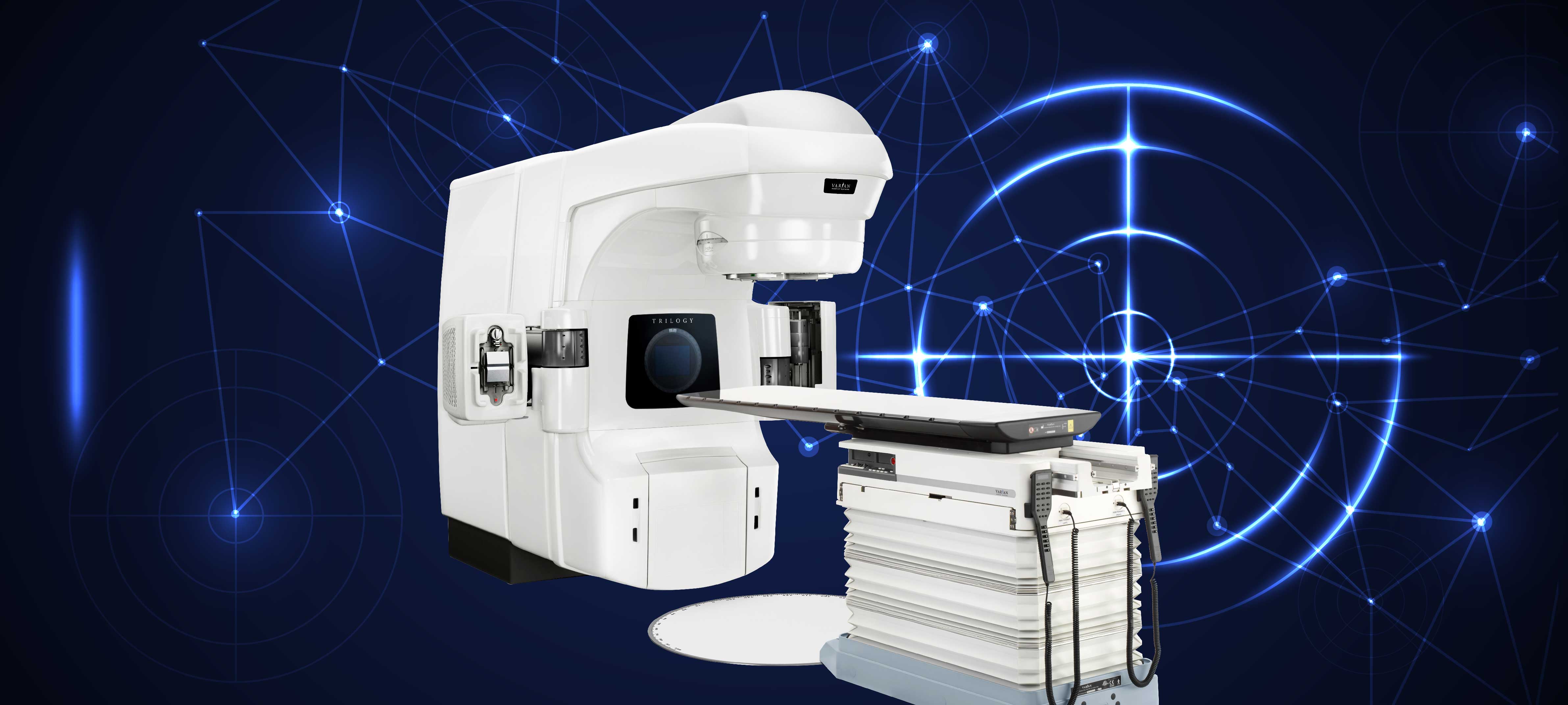
Radiotherapy is a matter of teamwork and requires a process. The patient needs to have a tomography scan for radiotherapy planning. While tomography is scanned, the patient is placed on a flat table and scanning is performed in a comfortable position, provided that the target area is included. On tomography scan, target organ and adjacent organs are identified in every slice in digital media. An accurate plan is made for each patient after radiotherapy dose, exposure of intact organs to how much dose and potential risks are calculated. Next, in the first day of radiotherapy, the patient is placed on the table at exactly the same position and radiotherapy plan is applied on the patient. At this stage, conformity of therapy plan is accurately verified on the patient in the digital media and each session is completed. Radiotherapy is applied for five days in weekdays and the therapy is skipped in weekends. It takes 10 to 15 minutes to verify therapy fields and 3 to 5 minutes to delivery radiation beams. Similar to roentgen scan, patient feels no pain while radiotherapy is applied; moreover, patient is monitored by technicians with camera at different angles.RADIOTHERAPY DEVICE IN OUR HOSPITAL AND ITS SPECIFICATIONSLinear Accelerator (LINAC) TRILOGY device, that includes 5 radiotherapy methods including CONFORMAL, IMRT, VMAT, SRS and IGRT, is used for diagnostic and therapeutic purposes.3D CONFORMAL3D CONFORMAL radiotherapy implies a therapy planning procedure where 3D tumor volume and critical organs are drawn using computed tomography images. In this modality, it is aimed to give the normal tissue the lowest possible dose while obtaining a dose that surrounds the tumor in a best possible way by giving a certain margin of target volume by multi-leaf collimators (Multi-leaf Collimator-MLC) of the linear accelerator.IMRTWith IMRT, intensity of X-ray beam is modulated. Thus, the intensity of the radiation in therapy area is adjusted and dose distribution is almost optimized. While high doses are applied to tumor, healthy tissues are conserved to the maximum extent.VMATVMAT (Volumetric Intensity Modulated Arc Therapy); it is a method of radiotherapy in which therapy is given in a very short period of time. In VMAT, duration of therapy is short relative to devices that do the same job. When radiation time of 2-3 minutes is added to imaging time that is less than 2 minutes, daily therapy is completed in 4 minutes totally in VMAT. While VMAT is performed, MLCs are moved according to positions of tumor and critical organs, and gantry and dose speeds are also changed during irradiation. Higher dose is more precisely targeted to the tumor using one or more than one arcs around the patient, while protection of sensitive organs is further maximized. With these features, VMAT can be defined as a simple, fast and effective radiotherapy method that hits the target precisely and correctly.
SRSCRS SRS technology enables punctuate radiation. With this method, punctuate radiation can be given to very small tumors that measure several millimeters in size. Therefore, while the tumor is applied high radiation dose, surrounding normal tissue is given lower amount of radiation.IGRTImage guided radiotherapy can be applied with IGRT. Image Guided Radio Therapy (IGRT) can be performed by using X-ray source on the remote control handles that are installed on the main body of our device, and reciprocal detector. With the help of IGRT feature, kV-kV image of the area of the patient, that will be treated, and Digitally Reconstructed Radiotherapy (DRR) images, those are taken by planning computer, can be evaluated in 2 dimensions before each therapy, and more importantly, Computed Tomography images can be obtained by turning robotic arms of the therapy device around the patients, and these images can be compared to reference images those are obtained by CT, that was scanned previously at simulation phase. Therefore, the patient can be included in therapy with same repeatability every day, and “set-up” uncertainties, those are originating from movements of organs, are minimized.
Doctors

Elnur SAHIBOV
M.D.
Related Patient Stories

Mr. Kenan applied to our Oncology Clinic with the diagnosis of Prostate Cancer
Mr. Kenan applied to our Oncology Clinic with the diagnosis of Prostate Cancer. A life system full of peace, abundance and health for Mr. Kenan, who survived cancer and regained his health after treatment.
Read Story
Orthopaedics and Traumatology
ORTHOPAEDICS AND TRAUMATOLOGY
Our experienced academic staff and specialist physicians, who benefit from vast opportunities of the modern technology, provide all patients with world-class medical and surgical treatments through a scientific approach.Our physicians examine congenital or developmental abnormalities of the musculoskeletal system through a multidisciplinary approach. Moreover, our center, that offers a healthcare service in a wide range of fields such as spinal surgery, hand surgery, bone tumors, hip and knee arthroplasty, sports injuries, fractures and dislocations, and pediatric orthopaedics, successfully performs diagnostic and treatment procedures of all musculoskeletal diseases, especially the diseases related to Orthopaedics and Traumatology. In general, the fields of study can be summarized as traumatology (musculoskeletal system disorders occurred due to hit-impact-accident), arthroplasty (joint replacement surgery), arthroscopic surgery (closed surgery procedures performed for intra-articular problems), hand surgery-microsurgery (including repair of severed organs through sutures), spinal diseases and surgery, pediatric orthopaedics (including congenital musculoskeletal disorders), musculoskeletal tumour surgery, and sports medicine.
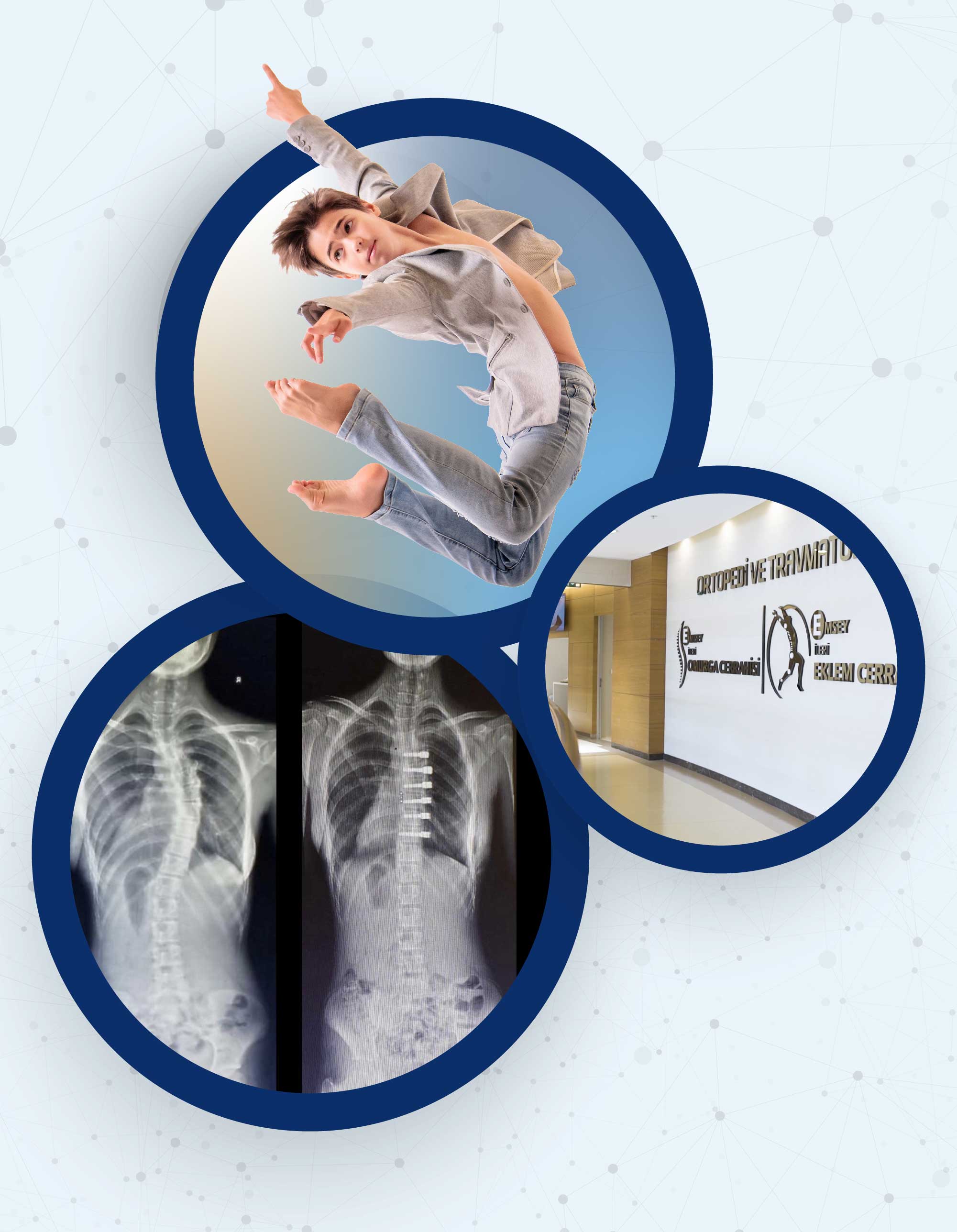
SPINAL DISEASES AND SURGERY
Spinal diseases and surgery is our another field of study. Spinal curvatures and fractures are the main parts of the treatment. Spinal curvature disorders may be congenital or developmental. Though spinal fractures usually develop after high-energy injuries, sometimes osteoporosis or tumour dissemination may have a role in development of spinal fractures. The nerves, that pass through the vertebrae and move from the brain to the locomotor system, increase the risks of this region. Diagnosis and treatment of spinal diseases are performed successfully in our hospital.
How is scoliosis treated?
There are many causes that may lead to scoliosis. However, different treatment methods have been developed for nearly 80 percent of patients who are diagnosed with scoliosis, in other words, spinal curvature. The methods that are commonly used for treatment of scoliosis in our day are divided into three groups as scoliosis exercises, scoliosis corset, and surgery for scoliosis. The most appropriate treatment method for scoliosis is determined after a detailed examination to be made by a specialist doctor. The treatment methods vary by person to person and the degree of spinal curvature.
Scoliosis in Adults
Scoliosis can be seen not only in children, but also in adults. However, scoliosis in adults is usually seen as it could not be diagnosed in childhood and has been brought into adulthood. Scoliosis may also be observed after the age of 50 due to spinal degeneration. Unlike scoliosis in childhood, the most common complaint of adults with scoliosis is pain. The pain is usually relieved with painkillers and physical treatment methods. As skeletal growth has ended, brace procedure with scoliosis corset is rarely performed. Surgical treatment may be required in some cases that are resistant to the relevant treatment methods.
Surgical Treatment
Surgical treatment is preferred if spinal curvature is advanced. Surgery should be considered if an individual’s curvatures on the back are more than 45 degrees or curvatures on the waist are more than 35 to 40 degrees. It is an extremely important surgery that requires a great attention. Therefore, surgery for scoliosis should be performed by an experienced and specialist doctor.
TRAUMATOLOGY
Traumatology can be defined as a sub-branch that deals with treatment of musculoskeletal system diseases caused by hits, impacts, falls and traffic accidents that are frequently encountered especially in our country. Even though fractures are the first thing to come to mind, cranial fractures are examined and treated by different departments. Facial fractures are treated by the department of Plastic-Reconstructive and Aesthetic Surgery and the department of Otorhinolaryngology Surgery. On the other hand, fractures of other cranial bones are treated by the department of Brain and Nerve Surgery. In addition, rib fractures that are frequently seen are treated primarily by the department of Thoracic Surgery. Fractures do not always develop after trauma. Injuries of the structures such as muscle-tendon-ligaments, that are generally called soft tissue injuries, are repaired and treated by the department of Traumatology. As we are a surgical department, surgical interventions are performed successfully by our team in cases where a surgical procedure is needed.
ARTHROSCOPIC SURGERY
Arthroscopic surgery can be defined as a procedure that is used in diagnosis and treatment of diseases inside a joint through a closed surgical method. Closed surgery refers to an intervention in which a pen-shaped camera is entered into the joint and the procedure is performed with pen-shaped tools from the other side. The most important advantage of the closed surgery is that it is a procedure performed without any surgical incision. The patient can return to her/his work earlier than an open surgery. Another advantage of this procedure is that it allows the doctor to make a detailed and functional examination of the synovial membrane, ligaments, cartilage structures, tendons and bone structures. Even though the interventions are most frequently performed for the knee and shoulder joints, arthroscopic interventions can also be performed for the elbow, wrist, hip and ankle joints. All arthroscopic interventions are performed successfully in our clinic.
ARTHROPLASTY
Arthroplasty can be shortly defined as joint replacement surgery that is performed with prosthesis. The word ‘’prosthesis’’ can be considered as the prostheses used in dentistry. Dental prostheses used in patients who have missing or highly deformed teeth replace the teeth and function. Similarly, prostheses are used in the treatment of cartilage degeneration that appears after the age of 40 in average or secondarily to any previous trauma. The prostheses replace the joints impaired. Though knee and hip replacement surgeries are usually more common, there are replacement procedures performed also for the shoulders, elbows, ankles, wrists and fingers. However, these prostheses should not be confused with the prostheses used to provide an appearance and function after any loss of a part of the body (amputation). For instance, the devices used externally to provide walking in a case of amputation of the leg are also called prosthesis. After the arthoplasty surgeries, the patient’s joint pain is removed and her/his mobility is increased. The prostheses can be permanent up to 15-20 years. These surgeries are performed successfully in our hospital.
MUSCULOSKELETAL TUMOURS
Diagnosis, treatment and follow-up of musculoskeletal tumours are made successfully in our clinic. The word ‘’tumour’’ refers to all kinds of cells that grow in the body. Moreover, tumours are divided into two groups as benign tumours and malignant tumours depending on their dissemination and ability to deteriorate the relevant structure. Musculoskeletal tumours can be basically classified as soft tissue tumours and bone tumours. In addition to surgical treatment, medication and radiation therapy are sometimes included in the relevant treatments.
PEDIATRIC ORTHOPAEDICS
As the name suggests, Pediatric Orthopaedics that is a sub-branch of medicine deals with musculoskeletal system diseases of children. However, it should not be forgotten that children are not miniature adults. Diagnosis, treatment and follow-up procedures of pediatric diseases are very different from the procedures performed for adults. Moreover, there are some interrelated disorders in which the department of pediatric orthopaedics can follow together with the departments of traumatology, spinal surgery and other sub-branches. However, when pediatric orthopaedics is considered, congenital gait abnormalities such as hip dislocation, that is a common disorder seen especially in our country, come to mind. Even though the incidence rate of hip dislocation has decreased gradually in recent years, it is still a problem. However, hip ultrasonography is a revolutionary development on this matter. Early diagnosis and nonsurgical treatments can yield very successful results. Ultrasonography of the hip is performed within the standard newborn screening procedure in our hospital.
HAND SURGERY – MICROSURGERY
Even though surgical suture procedure performed for severed parts of the body with microsurgical methods primarily comes to mind when hand surgery-microsurgery is considered, it can be said that hand surgery covers a wide range of areas from the fingertip to the shoulder. When microsurgery is considered, the procedure, in which the vascular and nerve structures that are almost invisible with the naked eye are sutured with very thin sutures and special suture tools under a microscope, should come to mind. Even though this intervention has become highly popular especially in the recent years, it is not a method that can be performed for every severed part of the body. Successful results can be achieved only if this procedure is performed for appropriate patients. It is very difficult to achieve successful results in patients with highly damaged vascular and nerve structures. Hand surgery should not be understood simply as a procedure in which the severed parts of the body are sutured. In hand surgery, all musculoskeletal injuries of the region between the hand and the shoulder are also treated. In addition, many examples that include bone fractures, nerve compressions and muscle-tendon tears in that region can be given. In our department, it is possible to successfully benefit from all opportunities of hand surgery and microsurgery.
SPORTS MEDICINE
Sports medicine, as the name suggests, is a sub-branch of medicine that deals with musculoskeletal injuries in athletes. Athletes may return to sport activities only after a true diagnosis, treatment and rapid rehabilitation program. Here, the most critical problem is the shortened time for return to sport. Some musculoskeletal disorders require a surgical treatment only in athletes. Return to sport is provided only through such procedure.
PROSTHESES (ARTHROPLASTY PROCEDURES)
Hip Replacement (Arthroplasty)
Replacement surgeries are also known as arthroplasty procedures. Hip replacement procedure is performed in cases where the hip joint is disrupted. Hip replacement surgery is necessary if the joint is disrupted due to arthritis of known or unknown origin, previous treatment performed for hip dislocation in childhood, hip-related fractures and dislocations, medication-related osteonecrosis of the hip joint, or previous Perthes disease in children.
Knee Replacement (Arthroplasty)
Knee replacement procedure is performed to correct the knee deformities caused by calcification of the knees and rheumatic diseases in elderly patients, fractures related to the knee joint, and sometimes the use of cortisone or similar cases that result in osteonecrosis that lead to a progressive destruction of the knee bones.
Doctors

Abdülhalim AKAR
M.D.

Medical Oncology
The Medical Oncology Department deals with Adult Oncology cases including Lung, Breast, Prostate, Gastrointestinal (Intestinal, Pancreatic, Stomach etc.), Thyroid, and Gynecological cancers. The entire spectrum of neoplastic diseases including benign and malignant cancers are treated at our Oncology Center. A multidisciplinary approach is used during the diagnostic and treatment stages to provide the best possible treatment in a patient specific approach. The latest Chemotherapy, Immunotherapy and Targeted Smart therapy protocols are some of the treatment modalities used at our clinic.
Oncology deals not only with diagnosis and treatment of all tumors and also all developments in this field. Many studies are conducted and new techniques are developed to ensure early diagnosis of various types of cancers. Computed tomography, ultrasound, nuclear magnetic resonance imaging and PET/CT are only some examples.
Chemotherapy uses potent medicines to destruct cancerous cells. The drugs are usually injected or administered by drop counter. This therapy may, sometimes, require staying at hospital.
Chemotherapy is accompanied by side effect, because drugs may also damage healthy cells. Hair loss, tiredness, feeling sick, vomiting and immunodeficiency can be observed. Sexual life of people can be also influenced. Patients may generate various reactions; all these reactions do not always develop simultaneously.
Treatment and follow-up are planned and applied for patients, who are diagnosed with cancer, at Department of Medical Oncology in our hospital. Relevant Surgeons and Radiation Oncologists cooperated in treatment and follow-up of these patients, if required. Advancements in treatment plans ensure that patients are followed up by Medical Oncologist, Chemotherapy Nurse, Radiologist, primary surgeon, a psychologist experienced in oncology and Dietetics and Nutrition Expert and treatment is personalized according to needs of patient. Our principle in treatment of patient is to play efficient role in resolving medical conditions, including but not limited to infections, renal failure, heart failure, nutritional disorders and so on, which are secondary to chemotherapy. Chemotherapy agents, which are intravenously infused, are prepared safely by an expert Chemotherapy Nurse in a special environment. At this outpatient clinic, patients are referred to inpatient settings for chemotherapy administrations, if required.
Diagnosis and treatment services are available for types of cancers written below at Department of Medical Oncology:
• Lung cancer
• Prostate Cancer
• Breast cancer
• Head & Neck cancer
• Stomach – bowel cancers
• Brain Tumors
• Pancreatic Cancer
• Liver cancers
• Gall bladder and biliary tract cancers
• Malignant mesenchymal tumors
• Kidney and urinary bladder cancers
• Melanomas and lymphomas
• Thyroid cancer
• Testicular Cancer
• Ovarian, endometrial and cervical cancers
Doctors
Abdallah TM SHBAIR
M.D.
Assoc. Prof. Dr.
Serap KAYA

Prof.
Emin Gökhan KANDEMİR
M.D.

Cardiovascular Surgery
In our hospital, modern minimally invasive cardiac surgery (with less damage) approaches are applied with the latest technology.
In our hospital, modern minimally invasive cardiac surgery (with less damage) approaches are applied with the latest technology.
MINIMALLY INVASIVE CARDIOVASCULAR SURGERY
In our hospital, modern minimally invasive cardiac surgery (with less damage) approaches are applied with the latest technology.
Axillary Heart Valve Surgeries
Commonly called "underarm or closed heart surgeries, scientific name is right anterolateral mini-thoracotomy, it is a 4 or 5 cm incision from the armpit, without cutting muscle and bone and making no incision to any other part of the body. The only difference between anterior surgery and armpit surgery is the entrance. The procedure performed inside is standard heart surgery. Also; With this method, heart surgeries are performed without cutting the anterior chest bone an average of 26-30 cm and opening it. None of your bones are cut.
In which heart problems, armpit surgery is preferred?
Since 1997; We use this approach in ASD (heart hole closure), mitral valve replacement and repair, tricuspid valve repair and aortic valve replacement. Also; Similar aesthetic methods are also available in coronary bypass surgeries.
What are its advantages?
- The risk of bleeding is low.
- The risk of infection is low.
- The recovery time is much faster.
- Even the patient himself cannot see the operation site.
- Aesthetically and psychologically, the patient seems to have not been operated.
- It does not cause sexual and social problems.
- After the operation, the patient can lie in the desired position.
- After the operation, the patient does not have to avoid the blows to the chest.
- Patient can drive a car when discharged and wear his seat belt.
- Can use his arms as if he had never had surgery.
- Can lift heavy.
It is an ideal method for young patients, especially those with risk factors such as obesity, diabetes, heart failure. It is easily performed for mitral and tricuspid valves in elderly patients, but it is not preferred in patients over 75-80 years of age, those with chronic lung disease and those who have previously undergone right chest lung surgery. In this group of patients, especially in aortic valve replacement, it is preferred to open a small front window in terms of aesthetics and less damage.
Can this method be applied to children and babies?
This method was first applied by Japanese scientists for heart holes in children. In other words, it can be applied in some heart surgeries such as heart holes in children and babies.
What should patients who prefer this method pay attention to?
They should prefer our physicians who have worked for years in University or Education, research hospitals on less damaged heart surgeries, which we call Minimal Invasive Surgery, and who have proven their experience or have training certificates by publishing the patients they operated with this method in national or international scientific journals and congresses.
Is it just as reliable as other methods and what is the risk?
One hundred percent reliable. The risks of anterior and lateral surgery are the same. When there is an inoperable situation, the surgeon instantly enlarges the area and switches to the normal entry method. Therefore, the entry method does not increase the risk of the surgery performed. Risk in heart surgery is associated with delayed surgery and heart strength.
Which method is more risk-free in second heart surgeries?
It is risky to open the patient from the front for a second time and the most ideal method for second and third mitral and tricuspid valve surgeries is to perform the surgery under the armpit. However, it is not applied for the second and third time in aortic valve surgery.
BYPASS SURGERY ON THE WORKING HEART (MINIMALLY INVASIVE)
When we say Heart Surgery, we all get scared and look for ways to get away from it. They take such remedies that sometimes we hope that the doctor or someone else will say, "You don't need surgery." But in vain… the risk increases a little more every day we run away and we may lose the chance of surgery. However, it should be the best to research "how can I get through this surgery lighter" Because in recent years, these surgeries can be performed by minimizing the intervention site we call "minimally invasive cardiac surgery" and without using a heart-lung machine, that is, without stopping the heart.
What advantages does this provide?
1. Reduction of surgical trauma and blood loss
It provides great comfort for the patient that the chest bone is not opened all along or not at all. It's like the difference between a stick of a needle and a large nail. The bleeding will be less due to the small amount of the opened area. In addition, postoperative pain is reduced.
2. Quick recovery and early discharge
The small wound heals quickly, especially if there is no bone and muscle incision, the patient gets up early. Patient can lie in the desired position on the bed and turn. Using his hands and arms, patient can meet his own daily needs. Patient can wear his seat belt, he does not have to avoid a blow to his chest.
3.Better cosmetic appearance
In these operations performed under the armpit, under the breast or under the breast, even the patient may not be able to see the operation site himself. Within a month, the intervention location becomes vague. It is preferred especially for young people, women and children.
4. Preventing Sexual and Social Problems
In open heart surgeries performed by stopping the heart and cutting the breastbone from beginning to end, many organs, especially the brain and heart, are affected by the heart-lung pump. Postoperative hair loss, personality changes, sexual reluctance and dysfunction may occur. In addition, sexual intercourse is prohibited until the breast bone is healed. However, none of these problems occur in aesthetic surgeries performed while the heart is working. The patient returns to his normal performance in a short time.
5. Low risk of infection,
The immune system is not damaged, the blood is not mechanically traumatized, and the blood cells remain intact. As a result of all these, the risk of infection and inflammation of the intervention site are very rare.
6. Reduction of brain, kidney and lung complications
When the heart-lung pump is not used, blood cells are not affected and complications such as brain damage, stroke, kidney failure, etc.
Due to these advantages, they are the most preferred surgical techniques today.
BUERGER'S DISEASE
Slight bruising on our feet or fingers or the wound on the nail can sometimes lead to the cut of our fingers. Usually we do not care much about these complaints and when we lose our finger we cannot believe it. Think of it first, your toe is cut off and later on your foot is cut off…. Your limb is gone!… And then the other foot, then the hands and the arm …… Here is this insidious disease, the result of which is terrible "Buerger"… No one should say I will not have it! Because although it is frequently seen in smokers and men, although the cause is still unknown, there is also familial transmission.
What would we do if we didn't have at least one of our feet that we didn't care about and that carried us for years?
The disease is usually seen in men aged 20-50 who smoke. Its symptoms are completely caused by blood circulation deficiencies that develop as a result of vascular narrowing or occlusion. Pain when walking on the calf, light hair loss, and above all, cold, cramping and bruising in our toes are the main complaints. If the bruising on the finger is delayed as a result of a small wound while cutting our fingernail or tightening of tight shoes, it is necessary to see a specialist without delay. Unfortunately, we use many ointments to heal these wounds, and as a result, inflammation accompanies. Those who cannot find the cure have to accept that the finger is amputated.
Although the patient loses a limb, he continues to smoke. However, stopping smoking can reduce the progression of the disease. Fingers and feet can be saved with very simple treatments such as simple abdominal surgeries (LOMBAR SYMPATECTOMY SURGERIES) and smooth muscle relaxants to heal the wound and enlarge the small-diameter arteries. As long as finger or foot ulcers and gangrene develop before the treatment comes. Fingers can also be involved in the disease at a rate of 30%. Hand treatment (THORACAL SYMPATECTOMY) is more successful.
CAROTID ARTERY SURGERIES
Atherosclerotic plaque creates a swelling in the carotid artery and prevents blood flow to the brain, and sometimes it also causes clotting, and as a result, stroke and paralysis may develop. Surgery to remove the inner layer of the carotid (carotid) so as to include the atherosclerotic plaque is called carotid endarterectomy. Carotid endarterectomy (removal of intravenous lime) surgery is the most effective treatment method known in this field, and it prevents the events such as stroke and paralysis that may develop as a result of carotid stenosis.
There are two jugular veins (carotid) on both sides of the neck that carry oxygenated blood from the heart. Approximately at the level of the chin, these veins are divided into two separate veins, internal and external. While the outer carotid gives the branches that nourish the face and scalp, the inner carotid directly enters the skull and feeds the brain. As the age progresses, atheroma plaques may occur on the inner surface and wall of cholesterol and fat circulating in the blood.
As the plaque grows, the vein begins to narrow and harden. This is called atherosclerosis. In the carotid, the plaque usually develops in the inner and outer carotid form where the vein divides into two and extends more to the inner carotid. Since the surface of the plate is uneven, it begins to be covered with blood clots over time. When this event progresses, the vessel is completely blocked and the blood flow to the brain decreases significantly, and this usually results in paralysis. In addition, the gradual swelling of the plaque may ultimately lead to cracking and the fragments in it breaking off the vessel wall and going to the brain, blocking the smaller vessels in the brain, and as a result, paralysis, blindness and speech disorders.
Endarterectomy surgery is performed for plaques that are narrow enough to cause a stroke threat.
Surgery indications (necessity)
Endarterectomy should be performed if there is severe stenosis (70% and above) that can cause the threat of paralysis, if temporary or permanent paralysis has caused blindness or speech disorders.Mild moderate paralysis in the last 6 months and 70% stenosis in the carotidIf there is a severe stenosis (more than 80%) in the carotid and does not have a complaint, the patient should be operated because the risk of paralysis is very high.Carotid endarterectomy is performed first, in the same session or after, depending on the patient's condition, in people who will undergo cardiac bypass surgery due to disease in their coronary vessels.
NON-SURGICAL VARICOSE TREATMENT
Today, especially the fearful dreams of women, but also evident in men, the disorders we call varicose veins occur as a result of the damage of the small valves in the veins that carry the dirty blood (poor in oxygen) to our heart against the gravity of the legs. Increased blood pressure within the vein can cause cosmetic problems, a feeling of heaviness in the legs, numbness, burning, edema and even ulcerated wounds. Although many factors are blamed in the formation of varicose veins, the cause of the disease has not been clearly determined yet. These factors are;
- Prolonged inactivity
- Standing constantly
- Excess weight
- Smoking
- Pregnancy and
- Genetic reasons ....
It is no longer a feared treatment method today. Because the treatments and surgeries performed without leaving scars can restore your legs. Among these; Foam therapy, whose Latin name is sclerotherapy, is one of the common methods used in the treatment of visible thin and medium capillaries. It is based on the principle of entering into the vein with thin-tipped needles, a foaming chemical when combined with blood adheres the vascular walls and eliminates the diseased area. It is a method that takes a short time to apply, can be performed under polyclinic conditions, gives immediate results, is painless and does not require anesthesia.
Excisional Laser: It is a trendy treatment method used in the treatment of superficial capillaries. The success rate is 80%. It especially destroys spider web images.
Endovenous Laser Ablation: It is a method based on the application of laser current with the help of a guidewire by inserting a small needle into the vein causing varicose veins. It is a very comfortable, easy-to-apply treatment method with very little bleeding and post-operative bruising. It is getting more widespread.
Radiofrequency ablation: Similar to laser ablation technique, it is a method applied by using radio waves instead of laser current into the vein. It has no superiority to laser treatment in terms of success rate.
In addition, some drugs can be used to heal the cells lining the inner surface of the veins and thus increase the strength of the veins. Diuretics and pain medication can be added to the treatment to relieve edema.
With these treatment methods, varicose veins are no longer a fearful dream.
However, early diagnosis and treatment are important for life comfort. Regardless of the stage, it is possible and easy to treat and apply treatment methods. Postponing and avoiding illness raise bigger problems. As a branch, it is within the area of work of cardiovascular surgery specialists. In particular, we recommend that individuals at risk and with symptoms of the disease consult physicians specialized in this area. It is gratifying to fight the disease with the treatment methods chosen and applied correctly in the hands of experts.
LASER TELENJECTASIA (spider web red spots) TREATMENT
When very thin capillaries under the skin expand, the image in the form of a red spider web is very disturbing, especially for women. These are not serious illnesses and do not cause discomfort. The latest technological treatment of this disturbing image is Laser application. These untreated cobweb structures spread rapidly.
It is a treatment performed by applying light carrying high heat energy to the vein. It is a method used in the treatment of capillary varicose veins. It is based on the principle that the intense laser light applied on the skin cooled with ice mold is held by the darker varicose and warms and disappears. The vessel wall and the blood cells in it, which are warmed up to 70-90 degrees, are destroyed and carried by the body and destroyed. It provides permanent treatment for the applied capillary varicose veins, that is, the treated vessel disappears forever.
Advantages
- Application is easy and painless, no drugs or narcosis required.
- It does not require rest, the patient can continue to work during the treatment.
- Does not leave permanent traces (scars are an important problem especially for women wearing skirts)
- It can be applied to any area including the face.
- If there are stains in the application area, they will heal.
- Leg hair may fall out or weaken as the hair roots may also be affected.
- It is not necessary to use bandages or compression stockings during and after the treatment.
HEART HEALTH IN CHILDHOOD
Prevention of heart disease, which is seen as a serious problem in later ages, starts from childhood. Heart diseases that are treated with the correct diagnosis in childhood can be prevented from becoming problems in the future.
Cardiac diseases seen in childhood are divided into three as "Congenital heart disorders, Rheumatic heart diseases and Rhythm and electrical conduction disorders". Congenital heart diseases are seen at a rate of 1%. The most common of these is ASD, known as inter auricular hole. These types of holes can be closed very easily by surgery. Especially, it is a great comfort for both the child and the parents that it can be performed with a small operation under the right armpit. The same method can often be applied in the treatment of heart valves in children. Long-acting penicillin treatment has decreased the frequency of valve diseases until the age of 30s. n addition to regular nutrition of our children, physical activity and sports have reduced the incidence of this type of heart disease.
HEART DISEASES IN CHILDHOOD
1. Congenital heart defects:
They are anomalies that occur as a result of the failure during the development of the baby in the mother's womb. These disorders, also known as heart holes, have many different forms. In addition, developmental disorders in the auricles and ventricles and coronary vessel anomalies can be found together or alone. This group of heart diseases is the most common.
2. Rheumatic Heart Diseases:
Common throat infections occur especially with streptococcal microbes known as beta. This group involves the heart valves and although it is more common in developing countries, it is still active in our country. It can be easily treated with careful follow-up and timely intervention.
3. Rhythm and electrical conduction disturbances:
These are the disturbances in the electrical stimulation and conduction system that make the heart work slow, pause and accelerate. It mostly manifests itself in school age and adult ages. Congenital heart anomalies can also be found with this ailment and this occurs as the cause. Sometimes there is no underlying cause. In addition, heart muscle inflammation and pericarditis occur as the underlying cause.
AORTA ANEURYSM
Aortic vessel rupture (dissection), which is among the cause of death in perhaps a quarter of high blood pressure patients every day, may be the cause of sudden death or may be experienced unconsciously and may appear in general screening tests.
It is difficult to know when this time bomb, which we are often unaware of, could explode. In fact, every person with high blood pressure, over the age of 50, smoking, overweight should definitely have a computerized tomographic angiography at least every 2 years or at least have an echocardiography. Because the expansion of the vein before this rupture may be a sign. This means to be operated before tearing starts and the risk is low.
Aortic rupture occurs in two ways. A tear that occurs between the walls of the vein that comes out of the aorta and the heart and carries blood to the whole body, but does not leak blood out of the vein, which requires urgent operation. Another form is that the wall of the main aortic vessel bursts completely and the blood comes out (into the chest, into the abdomen). In such a situation, patient cannot even be taken to the hospital, death is sudden and often the cause of death is unknown and is considered as a heart attack. This situation can only be detected accidentally with preliminary examinations and it is taken into operation before bursting out.
It is very important not to waste time for diagnosis. The risk of the operation increases according to the size of the tear. Especially if the aortic vein goes from the outlet to the area that includes the carotid artery, the risk is high, especially if it descends into the abdomen to include the kidney vessels, the risk of the operation is at the highest level.
Over the age of 45, smokers, high blood pressure patients, diabetics, cardiac patients should definitely not miss follow up visits
Doctors
Prof.
Süleyman ÖZKAN
M.D.

Pediatric Surgery
A child is not a little copy of an adult. Children have unique anatomic, physiological, metabolic and physiological characteristics. These characteristics greatly influence diagnosis-treatment approaches and surgical interventions to be made for children. Moreover, even the same disease is managed using very diverse modalities in children and adults. Therefore, the children should be operated on by surgeons, who are educated on diagnosis and treatment of pediatric diseases, namely pediatric surgeons.
Even if very minor surgeries are not properly performed in children, very severe complications are likely to occur. Pediatric surgery is a field of science that has developed to meet these requirements. This branch of medicine deals with diagnosis and management of congenital and acquired diseases in children that begin in the intrauterine life and persist to the end of adolescence.In our Pediatrics Surgery Clinic, congenital and acquired diseases are successfully managed in the light of actual and scientific facts for children ranging in age from 1 day to 16 years.Most of the surgeries performed in our clinic do not require a hospital admission and children can be discharged on the same day. Children stay in single rooms together with their mothers and relatives, if hospitalized. Neonates undergoing surgical treatment due to congenital conditions are monitored and treated by pediatricians and neonatal intensive care nurses at neonatal intensive care unit in both pre-and post-operative periods.
URODYNAMIC LABORATORY;
It is used for diagnosis and treatment of urinary system diseases. (Neurogenic bladder, spina bifida, voiding disorders)
ANORECTAL MANOMETRY LABORATORY;
It is used for diagnosis of constipation, fecal incontinence and congenital anal anomalies.
PEDIATRIC ENDOSCOPY UNIT;
It is used for diagnosis and treatment of diseases of stomach, small intestine and colon. In pediatric patients, imaging studies and surgical procedures are done for esophagus, stomach, small intestine and the entire colon.
NEONATAL DISEASES
After prenatal diagnosis is made, family is informed about the course of action and following procedures in post-natal period.
Common disorders, mostly congenital, for neonates (from birth to the first 30 days of life) are as follows:
Esophageal atresia, congenital and developmental anomalies of lungs leading to respiratory distress in children, diaphragmatic hernia, intestines into the chest cavity. Omphalocele and gastroschisis (extra-abdominal location of bowel secondary to atresia of abdominal wall). Congenital Intestinal Obstruction, anal atresia, congenital absence of rectum. Cysti hygroma, large cystic nodules/tumors in head&neck region, congenital aganglionic megacolon, absence or lack of nerves in distal segment of colon that prevents children from passing stools. Renal, urinary tract and urinary bladder disorders that can be identified in pre-natal or post-natal period. Every type of tumor in neonatal period, neonatal circumcision
PEDIATRIC UROLOGY
In urogenital system diseases in children, Pediatric Surgeons, Pediatric Nephrologists, Radiologists and Nuclear Medicine Specialists examine children and their test results. After treatment is planned, endoscopic, laparoscopic and open surgery interventions are carried out by Pediatric Surgery Clinic.
Hydronephrosis, hydroureteronephrosis, congenital urethral strictures, vesico-urethral reflux ( retrograde flow of urine from the bladder into the ureter, bladder dysfunctions, voiding disorders, urinary incontinence, penile anomalies, An abnormal direction of urine flow from the underside of the penis, (hypospadias), Epispadias, Penile curvature, Labored urination, revision of circumcision
OTHER COMMON DISEASES IN PEDIATRIC SURGERY
Surgical Diseases of Inguinal Region
Undescended testis
Inguinal hernia
Hydrocele
Varicocele
Torsion of testis
Gastrointestinal Disorders
Diagnosis and treatment of every kind of diseases resulting from intraabdominal organs are included.
Pediatricians and Pediatric Gastroenterologists closely cooperate with each other.
All the means of the modern medicine are successfully applied in surgical treatment for this group of diseases, including endoscopic and laparoscopic surgery.
Congenital anomalies of digestive system in which signs appear late.
Stricture of esophagus, secondary to caustic and corrosive materials
Stricture of esophagus in children, secondary to foreign body ingestion
Gastroesophageal reflux disease
Appendicitis, which is manifested by abdominal pain, should be considered for every child and can be harder to diagnose and results in further problem in younger children
Intestinal obstructions
Intestinal or gastric bleeding
Constipation or fecal incontinence
Hepatobiliary diseases
Treatment of Hepatobiliary diseases and congenital or acquired surgical diseases is done.
Cysts, abscess and tumors of liver
Congenital atresia, cystic dilatation or bile stone of bile ducts
Surgical diseases of pancreas
Cysts, sinuses, mass lesions, torticollis and enlarged lymph nodes in head&neck region
Thyroid diseases (goiter, cyst and tumors)
Congenital and acquired lung diseases
Congenital chest wall deformities, like pectus excavatum and pectus carinatum
Abscess and empyema secondary to pulmonary infections
Foreign body in respiratory tract
Emergencies like pneumothorax ( presence of air in the pleural cavity) and hemothorax ( presence of blood in the pleural space)
Trauma, traffic accident, fall and burn in children
Female reproductive system and genital diseases
Labial fusion or genital region adhesion in girls.
Imperforate hymen
Ovarian torsion
Ovarian cyst
Disorders of sex development, ambiguous genitalia
Doctors

Prof. M.D:
Zekariya İLÇE
Interventional Radiology
The Interventional Radiology department led by Prof. Dr Ahmet Kemal Fırat is high experienced in endovascular treatment modalities which are often required for certain neoplasms. These treatment options include endovascular embolization, deep tissue biopsies, chemotherapy catheter placement procedures and TARE/ TACE procedures. Our Cancer Center is able to offer patients minimally invasive procedures to increase patient comfort and enable complex cancer treatment options.
Our interventional radiology center, which is the therapeutic department of radiology, helps other clinical branches in many issues by applying the necessary treatment protocol with imaging devices such as angiography, computed tomography, ultrasonography, and magnetic resonance by using special needles, wires and catheters without creating a surgical incision.Interventional procedures that do not require surgery are performed under local and sedation anesthesia, making sure that the patient is comfortable, and drugs that relaxes the patient are given intravenously. This way, patients can be treated comfortably without feeling any pain. For these processes, the materials are specially designed and produced for each process and purpose, using mostly high-tech devices. These methods, which are painless and have very low risks, increase the quality of life of the patients, shorten the hospital stay, and patients are discharged on the day of the procedure or usually one day later.Special operations performed at our centerIn interventional radiology, biopsies can be taken from many organs such as liver, breast, lymph nodes, prostate; cysts and abscesses can be treated; clogged bile and urinary tracts can be opened; dialysis catheters can be inserted, and especially liver tumors can be treated by burning with special needles and drugs. Bubbles that cause brain hemorrhage or vascular diseases that cause stroke are also treated interventionally without opening the skull.Main operations performed in our center
- Tumor Marking
- Drainage (Liquid Discharge) Operations: Kidney and Urinary Tract Interventions, Abscess Drainage
- Biopsies
- Intravenous Catheters: Dialysis Catheter Insertion, Port Insertion,
- Picc-Line Catheter Installation
- Vascular Treatments: Vascular Occlusion Treatment,
- Diabetic Foot treatment
- Intracranial Vascular Aneurysm Treatment
- Bubbles that cause bleeding in the vessels feeding the brain can be treated by entering the vein. By using metallic stents and coils, the bubbles are occluded and bleeding is prevented.
- AVM Embolization
Doctors
Prof.
Ahmet Kemal FIRAT
M.D.
Radiology
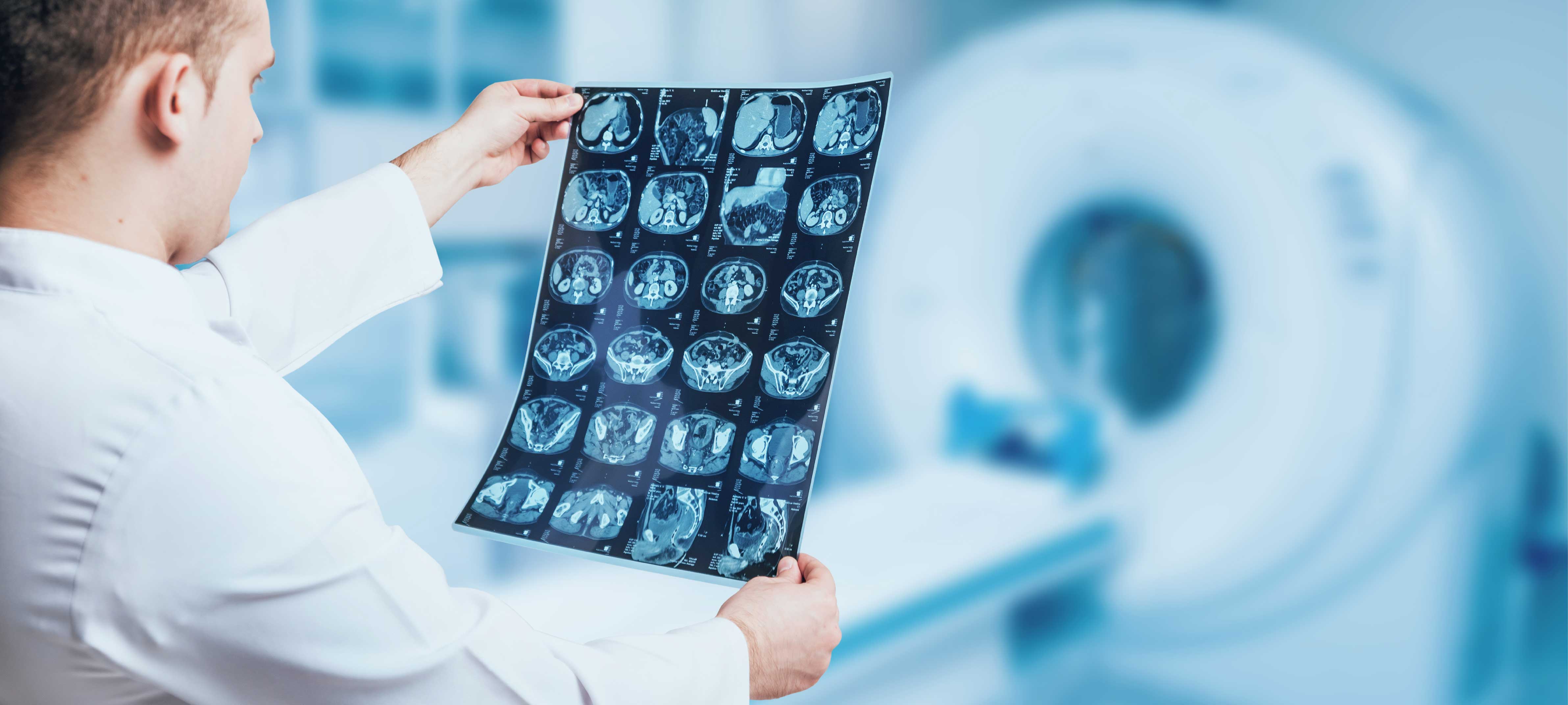
Radiology plays a critical role in the diagnostic and evaluation stages of oncology patients. Our Center is equipped with the latest radiological imaging modalities such as 1.5 Tesla and 3 Tesla Magnetic Resonance Imaging (MRI), Computed Tomography (CT), and Nuclear Medicine Imaging technology. Apart from the high quality technological devices and negligible waiting times, highly trained Radiology Specialists and Technical Assisting staff ensure reliable results.
Our Hospital possesses the most advanced state-of-the-art radiology devices and all the diagnostic and interventional radiological procedures are carried out.The Department of Radiology has experienced physician team and the technical equipment that can meet all diagnostic and therapeutic needs of both patients and doctors. Our personnel show ultimate attention during the procedures to ensure that the patients are comfortable.Radiologists have the knowledge and experience to perform not only diagnostic procedures such as general radiology, computed tomography, mammography, panoramic x-ray, digital fluoroscopy, magnetic resonance imaging, ultrasound, color Doppler ultrasound and angiography but also therapeutic interventional procedures. The department renders services 24/7 for patients and reports are quickly transferred to doctors to accelerate diagnosis and treatment.
ULTRASOUND (US) AND DOPPLER ULTRASOUND
All ultrasonographic studies are performed such as abdominal, pelvic, renal, thyroid, thoracic, breast, hip, eye and cranial ultrasounds. Moreover, services include transrectal ultrasound, transvaginal ultrasound and intraoperative ultrasound. In addition to these studies, ultrasound-guided fine needle aspiration and biopsies, fluid aspirations and catheter drainage procedures are performed.
Vascular structures and blood flow can be evaluated in detail with color Doppler ultrasound. Color Doppler ultrasound of arterial and venous system of upper and lower limbs, portal Doppler, Doppler ultrasound of carotid and vertebral arteries, orbital and scrotal Doppler, gynaecologic and obstetric Doppler, renal arterial Doppler and graft evaluation are also performed. These examinations are carried out by physicians who are highly experienced in this field. Unconditional patient satisfaction is one of the basic principles in the department; patients can schedule an appointment on the same day and waiting period for appointments is kept as short as possible.
All double contrast and biphasic examinations of gastrointestinal system are made and enterocylisis is performed to image the small intestine. Excretory urography, cystography, voiding cystourethrography and retrograde urography are amongst the genitourinary system studies. All hysterosalpingographies are performed under guidance of a gynaecologist and images are interpreted by radiologists. For fluoroscopic imaging, attention is paid to maximize quality of imaging, while minimizing the dose of radiation.
MAMMOGRAPHY
Mammography is a special examination performed with x-rays to diagnose breast cancer and other benign diseases of breast. Mammography services include screening tests for asymptomatic patients, diagnostic examinations for women with symptomatic breast diseases and stereotactic preoperative breast marking and consultations.
Technological specifications of the devices ensure optimal magnification and compression techniques with minimum radiation dose. The department provides accurate and fast diagnostic services for screening and diagnostic mammograms and galactography, ultrasound-guided breast marking and fine needle aspiration and biopsies are also performed successfully.
All images acquired by these devices are transmitted to PACS (Picture Archiving Communication Systems) and evaluated and stored by radiologists. As the system is integrated to the hospital information system (HIS), the results can be quickly reached by all outpatient and inpatient clinics and consultations can be made.
COMPUTED TOMOGRAPHY (CT)
CT can form detailed and cross-sectional image of any body part and can be used for diagnosis of many diseases, ranging from soft tissue diseases to fractures. It is frequently used in conditions where other imaging methods, such as direct x-ray and ultrasound, cannot provide certain results. Spiral or sequential scanning is performed for any body part, such as brain, vertebra, thorax, abdomen and osseous and pelvic structures. Moreover, CT-guided biopsy and interventional procedures can also be carried out to reduce the need of surgery and for histopathological diagnosis of lesions.
MAGNETIC RESONANCE IMAGING (MRI)
All neurologic examinations (brain, neck and vertebral column), vascular angiography (peripheral, intracranial, carotid and aorta), abdominal, thoracic and cardiac examinations, breast MRI, musculoskeletal system examinations, MRI cholangiopancreatography (MRCP), fetal MRI and functional MRI can be scanned. Functional MRI techniques include blood oxygenation level dependent (BOLD) imaging, diffusion and perfusion MRI, MRI spectroscopy and cerebrospinal fluid flow analysis. For the patients for whom definite diagnosis cannot be established with mammography and ultrasound, breast MRI can be canned to diagnose the breast cancer. MRCP ensures that bile ducts and pancreatic canals are non-invasively imaged and rapidly become a substitute for diagnostic ERCP studies. Radiology studies that require anesthesia administration can be performed under supervision of anaesthesiologist for patients in every age group in the department.
Doctors

İshak İKİZ
M.D.

Hüdaverdi KARADEMİR
M.D.
Prof.
Ahmet Kemal FIRAT
M.D.

Fatma Hülya CENGİZ
M.D.
Asuman YİĞİT
M.D.

Hüseyin ÇAĞIL
M.D.
Obesity Surgery
BARIATRIC SURGERY
Bariatric Surgery, also known as Weight Loss Surgery, is the common name given to a group of surgical procedures aimed at helping obese patients lose weight. These surgical procedures are among the most effective treatment methods for preventing the life-threatening complications associated with morbid obesity.
PREOPERATIVE PROCESS
SURGICAL PROCEDURE
Sleeve Gastrectomy
Gastric Bypass Surgery (Roux-en-Y Gastric Bypass)
Biliopancreatic Diversion with Duodenal Switch
Gastric Balloon
POSTOPERATIVE PROCESS
Doctors

Prof.
İlker Murat ARER
M.D.

Prof.
Tarık Zafer NURSAL
M.D.
Related Patient Stories

Rebecca, who had Obesity Surgery regained her health
Rebecca, who had Obesity Surgery regained her health.
Read Story
Nuclear Medicine
The Nuclear Medicine Department is involved in both diagnostic and treatment stages of cancer treatment. PET – CT scanning, Gallium 68 PSMA scans, Scintigraphy procedures and Iodine scans are some of the cutting-edge diagnostics performed at our clinic. With the help of these advanced diagnostic modalities, our teams are able to perform high – quality staging and grading of cancer patients. Early diagnosis is one of the corner stones of effective cancer therapy and our clinic offers negligible waiting times for these screening and diagnostic procedures. Furthermore, the Nuclear Medicine Department also offers advanced treatment modalities including Actinium, Technetium, Samarium and Radioactive Iodine treatment options.
TREATMENT OF LIVER CANCER WITH MICRO-SPHERE METHOD
Liver cancer is a prevalent type of cancer not only in our country but also in U.S. and European countries. Successful outcomes are obtained in management of liver cancer with microsphere – a new generation therapy – along with chemotherapy and medication therapies. It took attention of medicine community due to almost total absence of side effect and other advantageous features.What are characteristics of liver cancer?Liver cancers are addressed in 2 main groups. First group involves primary cancers that imply cancerous formations originating from hepatic cells, while second group is metastatic tumors that metastasize from cancers of other organs. Primary liver cancers also imply hepatocellular cancers that originate from native cells of liver.What is difference of micro-sphere methods in comparison to other methods?Micro-sphere method is a specific therapy. Chemotherapy and medications are systemic therapy methods. In other words, they do not absolutely target the lesion in liver. “Specific therapy” implies that there will be no systemic side effect, or in other words, patient will not encounter exhaustive side effects that occur after chemotherapy.What is incidence of liver cancer?Five hundred thousand to 1 million new primary cancer cases are diagnosed per annum worldwide. When metastatic cases are considered, pancreas and colon cancers are most common ones.Hepatic metastases account for 50 to 70 of pancreas and colon cancers. This is an extremely high rate. Another striking figure is about the fact that 20% of deaths due to colon cancers arises out of hepatic metastases. In this respect, both primary cancers and metastatic cancers are of remarkable clinical significance.How is treatment applied?Micros-sphere method is based on principle of using micro-spheres that measure 20 to 50 microns in size and are labeled with Yttrium-90 – a radioactive material that emits beta particles. Micro-spheres labeled with Yytrium-90 are directly injected to arteries that supply blood directly to liver. Thus, preparation accumulates only on tumor tissues located in the liver.Since they accumulate in tumor tissues and occlude capillaries that feed tumor tissues, it is also referred as “radio-embolization method”. Femoral artery is punctured and a catheter is inserted (similar to coronary angiography), and thus, drug is directly administered into feeding artery of liver.
What are treatment methods?
There are various treatment methods for liver cancers. The first one is surgical treatment, although not all patients are eligible for surgery. This indication is related with tumor content of liver. If tumor load is very high in liver or if there is more than one lesion, surgical treatment is contraindicated. Chemotherapy and specific drugs are therapeutic options for group of inoperable patients. However, primary cancers of liver are particularly resistant to therapy.Since metastatic lesions are usually multiple in nature, strong response to therapy methods mentioned above cannot be obtained. Therefore, radioembolization method is used that is recently introduced to clinical use. In radioembolization method, microspheres (20 to 50 microns in size) labeled with radioactive material are directly delivered to feeding artery of liver and thus, therapy is applied.What are advantages and disadvantages of the method?Being a local therapy and absence of exhaustive side effects that are observed in chemotherapy are most significant advantages of micro-sphere method. The second advantage is that this therapy method can be easily applied to patients who already received chemotherapy, chemoembolization therapy or surgical treatment. Briefly, micro-sphere method can be combined with other treatments. If therapy helps findings of cancer, the procedure can be repeated, if required.Which patients are eligible for micro-sphere method?Since this method primarily targets liver cancers, they are applied to both metastatic or hepatocellular cancers. However, this therapy is not a therapeutic option in this combined group of patient, if tumor load is high, or in other words, liver is largely infiltrated. If patient has a condition of liver failure, it will be useless again. Preliminary tests are analyzed to determine patients who are eligible for this method.How many patient received this therapy and what is success rate?To date, many patients received this therapy worldwide. This procedure has been widely used in Turkey approximately for 4 to 5 years. This specific method is applied in several healthcare facilities in our country. Approximately 200 patients received this therapy per annum. This implies that minimum 1000 patients are treated, to date, with this method in Turkey, but it has been very widely used in U.S. and European countries for 10 to 15 years.Neuro-endocrine tumors are rich in somatostatin receptors (SSTr).Group of tumors rich in SSTrGastroenteropancreatic tumors (carcinoid, gastrinoma, insulinoma, glucagonoma, VIPoma)Sympatho-adrenal system tumors (pheochromocytoma, paraganglioma, neuroblastoma, ganglioneuroma)Medullar thyroid carcinomaPituitary adenomaMerkel cell carcinomaSmall cell lung cancerBreast carcinomaMelanomaLymphomaProstate cancerNon-small cell lung cancerSarcomaRenal cell carcinomaDifferentiated thyroid carcinomaAstrocytomaMeningioma.GA68 DOTA PEPTIDE PET/CT SCAN IN NEUROENDOCRINE TUMORS (SOMATOSTATIN RECEPTOR IMAGING)Conventional imaging methods are characterized with very limited performance and low sensitivity in staging, re-staging and evaluating therapy response in neuroendocrine tumors. Since glucose metabolism is slow in neuroendocrine tumors, sensitivity of FDG PET/CT scan is low. Since SSTr is imaged with GA 68 DOTA Peptide PET/CT scan, molecular SSTr content of neuroendocrine tumor is demonstrated in high sensitivity with this method.INDICATIONS OF GA 68 DOTA PEPTIDE PET/CTInvestigating localization and metastatic focus of primary tumor (staging)
Determining presence of residue, recurrence or progression (re-staging)Determining indication of Lu-177 therapy in case of metastatic disease according to Ga68 peptide uptake.Response to therapy (surgery, radiotherapy, chemotherapy or Lu-177 Peptide therapy)Special precautions taken before or after imaging. This examination is absolutely contraindicated only for pregnant women. GA 68 DOTA Peptide PET/CT imaging is similar to FDG PET/CT scan in terms of imaging technique. The only difference is radiopharmaceutical agent used.
Lutetium (Lu-177) PEPTIDE THERAPY IN NEURO-ENDOCRINE TUMORSSince Lu177 is a radionuclide that emits beta particles, it penetrates into tissue by 1 to 3 mm. Based on this characteristic, it enables specific and internal radiotherapy approach. Positive lesions on Ga 68 DOTA Peptide PET/CT will point out uptake of Lu177-labelled peptide.Lu-177 peptide is indicated for management of all inoperable and/or metastatic neuroendocrine tumors that show positive SSTr content on Ca 68 DOTA Peptide PET/CT scan.Administration: Lu177-labelled peptide is diluted with isotonic sodium chloride (10-100 ml) and intravenously infused at 10 to 20 minutes. Patient should stay, minimum for one day, in a single room that is specifically licensed by Turkish Atomic Energy Authority regarding radiation safety. Therapy cycle periods: Lu therapy is usually given in 4 to 5 cycles at 6- to 8-week intervals.
What are side effects?
Acute side effects: Nausea, vomiting, abdominal pain.Nephrotoxicity: Nephrotoxicity is minimized, to the possible extent, with renoprotective therapies.Bone marrow toxicity: It is a reversible condition that develops in 2-5% of patients especially if patients have diffuse bone metastasis.Efficiency of TreatmentLu177 have been used worldwide in management of neuroendocrine tumors approximately for one decade. This therapy has been available in particular healthcare facilities in our country approximately for 3 years. Literature data demonstrated that Lu-177 peptide prolongs survival, improves symptoms, restores biochemical markers into normal range and contributes to enhancement of quality of life in patients with SSTr-positive neuroendocrine tumor.
GALLIUM (68Ga)-LABELED PSMA PET IN PROSTATE CANCER
Prostate cancer is the second most common type of cancer in male subjects and sixth most common disease among all causes of death. Similar to all other cancer, it is very important to determine best therapy method timely in prostate cancers.Recently, total body is scanned and metastatic status of prostate cancer can be determined with Gallium (68Ga)-labeled PSMA PET scan. PSMA is a substance that is totally specific to prostate gland. When this substance is used in Galium (68Ga) PET scan, uptake is observed only in regions which are involved by prostate cancer.Current status of disease is revealed out with Galium(68Ga)-labeled PSMA PET scan and thus, best therapy method is selected.
Doctors

Yusuf Demir
M.D.
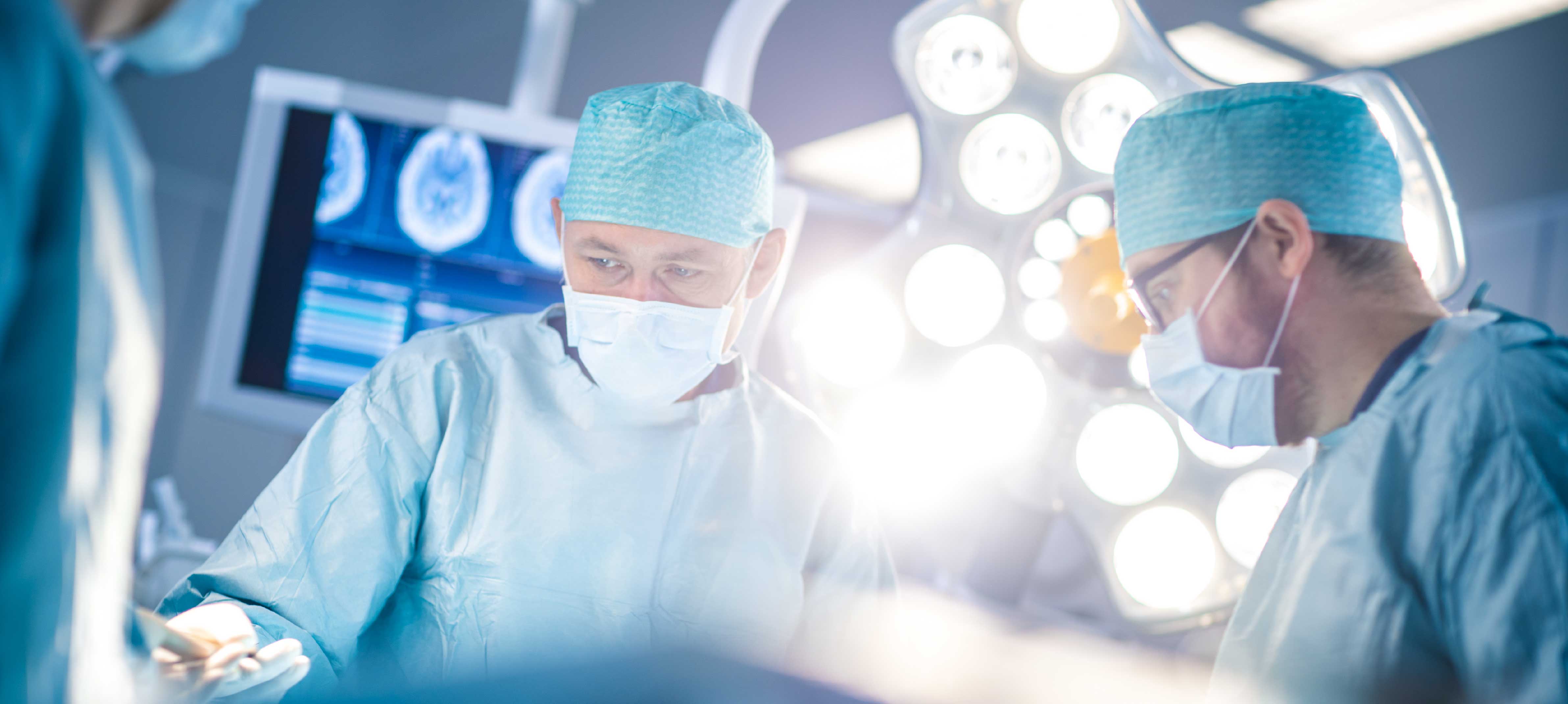
Oncologic Surgery
Apart from medical and radiotherapy approaches, our cancer center is equipped with experienced surgeons from all necessary branches to offer patients with surgical options during the course of cancer treatment. Oncoplastic surgery, Tumor Excision, Oncogynecologic, Urology, Head and Neck, Neurosurgery, Pediatric Surgery and General Surgery departments all participate in the Tumor Council to evaluate cases for the possibility of surgical intervention when necessary. Our center is suitable for complex or complicated cases requiring a multi-disciplinary approach consisting of a mix of multiple treatment modalities.
Related Patient Stories

Bosnian Sacira recovered after Oncology Treatment
Sacira Cengic, who came to our hospital from Bosnia, applied to our Oncology Clinic for cancer surgery. We wish a life full of peace and health to Ms. Sacira, who defeated cancer and regained her health after her treatment. Early diagnosis and correct treatment are important in cancer. Do not delay your health.
Read Story
Neurosurgery
Neurosurgery
Surgical treatment for diseases of the nervous system and OF the structures that surround the nervous system is an extremely sensitive and important issue. Therefore, searching a well-equipped and experienced centre with a good team is often of vital importance.In the field of the brain, spine, spinal cord and nerve surgery that is highly complicated and within a broad range, ‘’Neurosurgery Department’’ in our hospital has carried out its procedures in collaboration with the physicians and surgeons who are experienced in the fields of Neurology, Neuroradiology, Neuroanesthesia – Intensive Care and Psychiatry. ‘’Neurosurgery Department’’ in our hospital has been designed to provide 24 hour-service with the latest technological opportunities in surgical treatments of the brain, spine, spinal cord and nerve diseases in newborns, children and adults.In the intensive care unit that is managed by the automation system, patients are followed up meticulously in the pre-operative and post-operative periods, and their treatments are regulated coordinately with other related branches. Thus, all necessary treatments can be provided even in the most problematic patients in our hospital without any need for another center. In addition, Angiography, Computed Tomography (CT) and Magnetic Resonance Imaging (MRI) procedures can be performed for 24 hours. Our operating rooms have the capacity to provide service in the following procedures at any time of the day with all kinds of neurosurgical devices (microsurgery, endoscopy, CUSA etc.), that are made possible by the latest technological developments, and technical equipment support.Angiography, Computed Tomography (CT) and Magnetic Resonance Imaging (MRI) procedures can be performed for 24 hours.
ENDOSCOPIC DISCECTOMY
Full Endoscopic Lumbar Discectomy as the New Golden StandardWhen surgery is needed for a herniated disc?Surgical treatment should be considered if the patient cannot benefit from the conservative treatment methods called physical treatment, medication therapy and similar treatments, or if there are progressive neurologic findings such as foot drop, urologic problems and insistent pain despite the treatment.
What are the current treatment methods for herniated disc?In our day, there are two surgical methods used in the treatment of herniated disc. These are the posterior open method and the percutaneous method made through the skin. Microscopic microsurgery has been regarded as the golden standard of this procedure until now. Open microdiscectomy has come into use at the beginning of the 20th century. The excitement experienced by endoscopy in our day was similarly experienced when microscopic discectomy has come into use with the development of the microscope towards the end of the 1970s. It was very important to perform this procedure without harming the stability of the mobility called the stabilization of the spine. Microscopic microsurgery system is now an invasive method when it is compared to the endoscopic method. It should be accepted that endoscopic discectomy is the new GOLDEN STANDARD.
What are the methods used in endoscopic discectomy procedure?
There are two methods used in endoscopic discectomy procedure. One of the methods is the interlaminar approach. There is a bone structure called lamina behind the spine, and a yellow ligament called ligamentum flavum is seen between the lamina and the spine. The yellow ligament is reached with a small incision (4 mm-5 mm) made through the skin, and the whole hernia can be discharged. The other method is the transforaminal approach.
Can this surgery be performed with local anesthesia?
Though it is said that this surgery can be performed with local anesthesia, it is necessary to provide patient comfort without pain because this procedure is performed to relieve the patient’s pain. Naturally, this surgery should not be performed while the patient is awake.The patient needs to stay in hospital overnight. If the surgery is performed in the morning, it is possible to discharge the patient from the hospital in the afternoon or in the evening. Depending on her/his general condition and age, the patient can begin to walk after half an hour or two hours following the procedure. However, we keep the patient wait for a while in order that s/he can regain her/his consciousness after anesthesia and feel more comfortable. Moreover, this procedure can be performed more easily because the size of the inner tear is not increased. The procedure also reduces the risk of recurrence of the disc.
EPILEPSY SURGERY
Microsurgical resection in lesion-related epilepsies, and placement of vagus nerve stimulator (VNS) in resistant and non-lesional epilepsies.
PERIPHERAL NERVE SURGERY
Peripheral nerve anastomosis and nerve repair with microsurgical technique after trauma, and surgery for entrapment syndromes (carpal tunnel syndrome or ulnar tunnel syndrome etc.)
TREATMENT FOR INTRACEREBRAL CYSTS
Fenestration of arachnoid cysts (Endoscopic technique and microsurgical technique)
FUNCTIONAL NEUROSURGERY
Stereotactic biopsy, deep brain stimulation (DBS), implantation of a neurostimulator also known as brain pacemaker in patients with movement disorders and Parkinson’s disease, tremor rehabilitation, hemifacial spasm and trigeminal neuralgia rehabilitation (balloon compression, radiofrequency and microvascular decompression)
SURGICAL TREATMENT FOR CONGENITAL ABNORMALITIES OF THE SKULL
All craniosynostosis (a condition in which the sutures between the skull bones close too early) and craniofacial (deformities of the head and facial bones) surgery interventions that require an advanced experience, cranioplasty surgeries, and surgical interventions for the skull lesions (skull tumours, cysts etc.)
NEUROENDOSCOPY AND HYDROCEPHALIA TREATMENT
Endoscopic third ventriculostomy (ETV), ventriculoperitoneal, ventriculoatrial and lumboperitoneal shunt surgeries
VASCULAR SURGERY DISEASES OF THE BRAIN AND SPINAL CORD
Surgical interventions depending on vascular diseases of the central nervous system such as intracerebral hematoma, aneurysm, anteriovenous malformation (AVM), cavernoma and cavernous angioma
NEURO-ONCOLOGY
Surgical treatment of all kinds of brain and spinal cord tumours, glial tumours, meningiomas, pituitary tumours (including minimally invasive endoscopic endonasal interventions), cerebellopontine angle tumours, posterior fossa tumours, brain stem, pineal region, skull base, extramedullary and intramedullary spinal tumours.
SPINE AND SPINAL CORD SURGERIES
Cervical, lumbar and thoracic discectomies, vertebroplasty, kyphoplasty, spinal stenosis, stabilization (instrumentation) surgeries, spinal trauma (spine-spinal cord injuries), spine and spinal cord tumours, all kinds of congenital abnormalities of the spine and spinal cord, meningocele, meningomyelocele, tethered spinal cord syndrome, spinal cord cysts, scoliosis etc.
TRAUMA
Injuries of the skull, brain, spine, spinal cord and nerves, epidural and subdural hematomas, compression fractures.

General Surgery
Thanks to the depth of staff with different areas of interest, pioneering applications are carried out in gastrointestinal system (digestive system) surgery and hepato-pancreaticobiliary (liver, pancreas and biliary tract) surgery, especially in minimally invasive surgery, emergency surgery and endocrine (breast, goitre gland and adrenal gland) surgery. All interventions in colon and rectum (large intestine) diseases are performed using the most up-to-date technologies. In particular, a wide range of treatments are offered to our people in anal (rectal) region disorders such as haemorrhoids (haemorrhoids), fistulas or fissures (cracks).The disease grouping that can be examined, investigated and treated by the General Surgery Department is as follows:
• Thyroid Gland Diseases (Goitre, Hyperthyroidism)
• Breast Cancer and Benign Diseases of the Breast
• Oesophageal Cancer and Benign Diseases
• Anorectal Diseases: Tumours, Haemorrhoids, Anal Fissure, Anal Fistula
• Gallbladder and Duct Stones and Tumours
• Abdominal Wall and Inguinal Hernias, Postoperative Hernias
• Trauma and Emergency Surgery
• Stomach Cancer
• Hemorrhoids, Anal Fissure, Anal Fistula and Ingrown Hair Surgeries with Laser
• Surgical Diseases of the Small Intestine, Intestinal Obstructions
• Colon - Rectum Tumours, Inflammatory Diseases
• Benign and Malignant Tumours and Cysts of the Liver
• Obstructive Jaundice
• Pancreatic Cysts and Tumours
• Acute and Chronic Pancreatitis
• Surgical Diseases of the Spleen
• Various Soft Tissue Infections
• Laparoscopic Surgeries
• Obesity
• Diabetes Surgery
Laparoscopy is the observation of the abdomen using an optical device. This device is inserted into the abdomen through a small 1 cm incision below the navel. By illuminating the inside of the abdomen, it allows direct observation of diseases or problems involving the uterus, ovaries and tubes and, if necessary, simultaneous treatment with auxiliary instruments inserted through 3 - 5 mm holes in the lower abdomen. In laparoscopic surgery, operations are performed by means of a micro-camera inserted through several small holes in the abdominal wall, which is viewed on a screen.The Most Important Advantages of Laparoscopic Surgeries are as follows:
• There is very little bleeding during the operation.
• A very small scar remains.
• There is very little pain after the operation.
• Since there are no large incisions, the infection rate is lower.
• Since recovery is quicker, loss of work capacity is minimised.
• Hospital stay is short (maximum 1 day).
• It prevents intestinal adhesion due to surgery in the long term.
Doctors
Prof.
İlker Murat ARER
M.D.
Prof.
Tarık Zafer NURSAL
M.D.

Endocrine and Breast Surgery
The diseases dealt by endocrinology and metabolism diseases department are very common and their prevalence is ever increasing in our country, as is the case with around the world. Such diseases are very prevalent and endocrinology and metabolism diseases department deals with a very wide range of diseases that involves many systems and organs.
What is Endocrine System?
Endocrine system is formed by internal secretory glands.Internal secretory glands synthesize and secrete hormones. Hormones regulate various activities in our body. Certain types of hormones regulate reproduction, metabolism, growth and development. Hormones also control our reaction to the environment and help supply of energy and nutrition sufficient to maintain functions of our body. Hypothalamus, pituitary gland, thyroid, parathyroid, pancreas, ovaries and testicles, adrenal glands, fat tissue as well as endothelium (cells lining innermost wall of vessels) are secretory glands that form the endocrine system.
Who are Endocrinologists?
Endocrinologist is a specialist dealing with the endocrine system. Endocrinologists diagnose diseases that affect the endocrine glands. They are trained to treat various diseases which are generally complex and affect several systems in our body. Your primary physician refers you to an endocrinologist when you experience a problem related to the endocrine system.
What Kind of Medical Education Do Endocrinologists Study?
Endocrinologists study internal medicine for four or five years following 6 years of medical education. They spend further three years to learn how to diagnose and treat hormonal problems. It takes more than 13 years in total to be an endocrinologist.
What Do Endocrinologist Deal With?
Endocrinologists are trained to diagnose and rehabilitate hormone imbalance and related problems. They handle numerous types of cases.
- Thyroid:
- Thyroid nodules (ultrasound-guided thin needle aspiration biopsy specimen obtained to evaluate the condition (TFNAB))
- Thyroid cancers
- Thyroiditis
- Thyrotoxicosis
- Hyperthyroidism (Graves Disease, Toxic Adenoma, Toxic Multinodular Goiter, Thyroid Storm)
- Hypothyroidism (Primary Hypothyroidism, Central Hypothyroidism, Congenital Hypothyroidism, Myxedema Coma)
- Goiter, iodine metabolism
- Parathyroid Diseases
- Hyperparathyroidsm
- Hypoparathyroidism
- Diabetes Mellitus (moreover, Insulin Pump Treatments):
- Hypoglycemia (Low Blood Glucose)
- Hypertension (High Blood Pressure)
- Disorders of Lipid Metabolism
- Metabolic Disorders
- Over- or under-production of hormones
- Osteoporosis (bone loss)
- Bone Tissue Diseases
- Osteomalacia
- Paget’s disease
- Adrenal gland diseases
- Adrenal gland adenomas
- Incidentaloma
- Cushing syndrome
- Addison’s disease
- Adrenal crisis
- Mineralcorticoids and related abnormalities (aldosteronism)
- Pheochromocytoma
- Multiple endocrine system diseases
- Tumors of pituitary gland
- Acromegaly
- Prolactinoma
- Non-functional pituitary adenoma
- Infertility
- Growth retardation (short posture).
- Tumors of secretory glands
- Obesity
- Metabolic syndrome and its treatment
- Endocrinologists may also order certain tests to examine how endocrine glands function.
Doctors
Prof.
Tarık Zafer NURSAL
M.D.
Prof.
İlker Murat ARER
M.D.
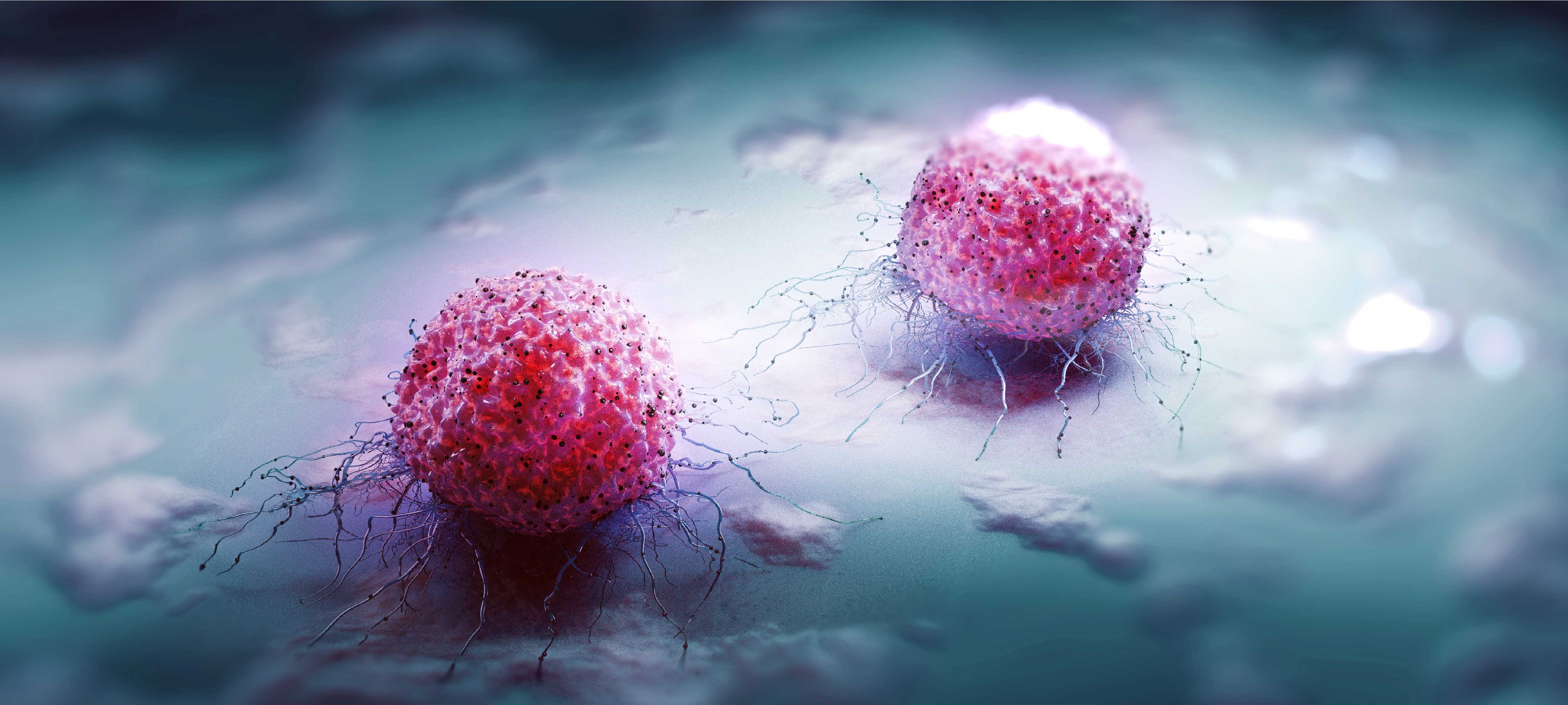
Hematology
- Anemia (Red blood cell deficiency, anemia)
- Leukopenia (White blood cell deficiency)
- Thrombocytopenia (Platelet deficiency)
- Polycythemia (Red blood cell excess, erythrocytosis)
- Leukocytosis (White blood cell excess)
- Thrombocytosis (Excess platelet)
- Sickle cell anemia: Anemia caused by a structural defect caused by a genetic mutation in hemoglobin molecules in red blood cells.
- Aplastic anemia: It is a rare, serious type of bone marrow failure that occurs when the bone marrow cannot make enough new blood cells.
- Autoimmune hemolytic anemia: It is a type of anemia that occurs when red blood cells are destroyed by the body's own immune system.
- Leukemia
- Lymphoma
- Multiple Myeloma
- Myelodysplastic syndrome (MDS)
2- Hematological diseases originating from platelets
- Hemophilia
3- Platelet function disorders
- Von Willebrand's disease
- Primary thrombocytosis
- Simple blood count
- Microscopic examination of blood
- Molecular biological tests
- Flow cytometry
What is Bone Marrow Transplantation?
- The “stem cells” found in the bone marrow are responsible for the production of blood cells in the body. In cases where the bone marrow is damaged or lost its function, the reintroduction of new “stem cells” obtained through various means into the body through the vein is called “bone marrow transplant”.
- The person from whom the stem cells are taken is called a donor/donor, and bone marrow transplants are classified according to the donor. Namely;
- Autologous transplant:
- The donor is the patient himself. Stem cells taken from the patient are frozen and transplanted back to him after certain procedures. The aim here is to reset the bone marrow damaged by high-dose chemotherapy and/or radiotherapy to be given for treatment. Multiple myeloma, Hodgkin lymphoma, non-Hodgkin lymphoma and acute myeloid leukemia are among the diseases that autologous transplantation can be performed.
Doctors
Prof.
Serdar Bedii OMAY
M.D.

Cardiology
The experienced staff in Cardiology Department of Emsey Hospital diagnoses many rare and complex heart pathologies as well as common cardiac diseases and plans treatment in the light of up-to-date scientific data.
The experienced staff in Cardiology Department of Emsey Hospital diagnoses many rare and complex heart pathologies as well as common cardiac diseases and plans treatment in the light of up-to-date scientific data.Our hospital follows up especially heart valve diseases, coronary artery diseases, congenital heart diseases, hypertrophic cardiomyopathies, heart failure and rhythm disorders (arrhythmias) with multidisciplinary approach and state-of-the-art techniques and technologies. Our cardiologists cooperate with the specialists of relevant branches, such as cardiovascular surgery team, endocrinologists, internists, pathologists, dietitians and physiotherapists, if required. This teamwork is “patient-oriented” and the patient is informed about possible treatment options and the most appropriate treatment is decided.
In this sense, every kind of interventional cardiac procedures (angiography, invasive treatment of coronary and peripheral arteries), pacemaker implantation and non-invasive diagnostic procedures ( rhythm Holter, ambulatory blood pressure monitoring, echocardiography laboratory, cardiac stress test, Thallium stress test) is performed successfully in the Department of Diagnostic Cardiology.
Moreover, healthy individuals are informed and educated on cardiac health and risk factors of heart diseases within the scope of preventive medicine
Diseases diagnosed and treated in Cardiology Department
- Myocardial Infarction (heart attack)
- Blockages of coronary artery
- Heart Failure
- Cardiac Rhythm and Conduction Disorders
- Heart Valve Diseases
- Peripheral Vascular Diseases
- Aortic Diseases
- Hypertension
- Hypercholesterolemia
- Congenital Heart Diseases
- Disease Risk and Screening
- All emergency procedures
- Invasive Procedures
- Myocardial Scintigraphy
- Transesophageal echocardiography
- Cardiac catheterization and coronary angiography
- Heart scintigraphy
- Invasive treatments (Coronary angioplasty, stent implantation, atherectomy etc.)
Electrophysiology Laboratory is used to diagnose and treat heart rhythm disorders. The problems identified there are eliminated with appropriate treatment options, like pace-maker implantation. Permanent pace-maker implantation is preferred in case of life threateningly slow heart rate. Pace-maker can also be implanted when the heart rate is significantly high.
Doctors

Mohamdou Ould TIJANI
M.D.

Assoc. Prof.
Selim TOPCU
M.D.

Pathology and Genetics
Biopsy results are generally regarded as the golden standard of diagnosis. Our hospital is equipped with a dedicated pathology lab capable of frozen section studies along with conventional and advanced pathological studies including immunohistochemical staining, immunofluorescence approaches and advanced genetic studies. Pathology Biopsy results form the basis of diagnosis and advanced genetic and hormone testing allow for patient-specific treatment approaches a such as immunotherapy and targeted smart therapy protocols.
Pathology literally means the science of disease. It examines the changes that occur at the cellular level in organs as a result of diseases. Tissue and cell samples are examined, and when necessary, they are examined by using techniques such as histo (cyto) chemistry, immune histo (cyto) chemistry, immunofluorescence, and FISH, in addition to the usual methods. Pathology diagnoses diseases by the appearance of cells under the microscope.In a pathology laboratory, cervicovaginal smear, fine-needle aspirations, tru-cut biopsies, small biopsies, excisional biopsies and operation materials are performed in detail, and if necessary, accompanied by USG or CT. In the cervicovaginal smear examination, liquid-based smear technique is used.In the examination of biopsy and operation materials, especially in tumor cases, it is recommended to perform advanced staining studies, such as histochemical and immune histochemical studies, if necessary.Emsey Hospital Pathology unit is in contact with all branches because of the multidisciplinary working principles of our hospital.
Doctors
Prof.
Hayreddin YEKELER
M.D.
Advanced Spinal Surgery
- Spinal Deformity
- Degenerative Diseases of the Spine
- Spine Injuries
- Tumoral Lesions in the Spine
- Infections of the Spine
- Decompression (Relief of Compression)
- Stabilization (Fixation)
- Generally refers to the procedures for restructuring and stabilizing the static balance of the spine, which is disrupted by traumatic, degenerative, and inflammatory diseases to make it balanced and stable again. Stabilization can be achieved with different surgical fixation methods using different materials. Lumbar shift (spondylolisthesis) is a good example of spinal disorders in which stabilization is frequently used. Spinal fusion surgeries are specialized stabilization methods where two or more bones in the spine are permanently connected to each other.
- Reconstruction
- Discectomy and Microdiscectomy
- Disc Prosthesis Applications
- Laminectomy, Laminoplasty
- Corpectomy
- Vertebroplasty
What is scoliosis?
- Can be defined as the right or left curvature of the spine when viewed from the front or back. However, it is actually a more complex deformity that affects the entire spine. In addition to moving sideways, the vertebrae also rotate in three dimensions, both upwards and downwards, and inwards and outwards.
- Idiopathic Scoliosis (Cause Unknown Scoliosis)
- Congenital Scoliosis
- Neuromuscular Scoliosis
- Functional Scoliosis
- Curvature and hump of the spine
- Difference in hip and shoulder levels
- Asymmetric appearance of body curves
- Asymmetric appearance between the body and arms
- Disproportionate appearance of the ribs
- Asymmetry of the chest
- Deepening of the lumbar curvature
- One scapula being more protruding than the other
- One shoulder being forward during walking
- One leg feeling longer than the other when walking
- Back and/or waist pain
- Shortness of breath
- Clothes that do not fit the body properly, do not fit properly
- Mild scoliosis (10-20 degrees of curvature)
- Moderate scoliosis (20-40 degrees of curvature)
- Severe scoliosis (>40 degrees of curvature)
- Usually involves radiological imaging and examination at certain intervals for curvatures smaller than 20-25 degrees. Regular exercise should be performed during this period. During monitoring, treatment options are evaluated based on the progression of scoliosis and are explained to the patient in detail by the physician.
After general scoliosis surgery, under normal conditions, 3-5 days of hospital follow-up may be required. Catheter is used for pain control for the patient's comfort during the first 24 hours. For patients who are standing up on the first day, removal of the chest tubes used in tethering surgeries can sometimes take 2-3 days. The full schedule for returning to work and full capacity sports activities is determined by the doctor during follow-up appointments, but it typically occurs within 1.5-2 months.
- (Also known as gibbosity, hunchback, or round back) is the name given to the spinal deformity where the normal outward curve is excessively increased when viewed from the side of the body.
- First and only "spine excellence center" certification in Asia and Turkey
- An academic team that leads the way in spine surgery in the medical literature
- Innovative and multidisciplinary approach: Scientific council in all fields related to spine health, such as spine surgery, orthopedics and traumatology, brain and neurosurgery, physical therapy and rehabilitation, neurology, rheumatology, and algology.
- Holistic treatment approach: Providing psychological, nutrition and diet, and rehabilitation support for patients under one center during postoperative process.
- Experienced healthcare staff in preoperative and postoperative preparation and care.
- 24/7 uninterrupted service throughout all diagnosis and treatment processes.
Doctors
Abdülhalim AKAR
M.D.
-

HEMOTOLOGY CENTER
-
GENERAL SURGERY
-
STEM CELL THERAPY CENTER
-
ADVENCED SPINE AND ORTHOPEDICS
-
UROLOGY CENTER
-
ONCOLOGY CENTER
-
CARDIOLOGY CENTER
-
EYE CENTER
-
BRAIN AND NERVE SURGERY
-
CARDIOVASCULAR SURGEON